. 2009 Jul 15;331(2):326-38.
doi: 10.1016/j.ydbio.2009.05.550. Epub 2009 May 19.

Persistence of histone H2AX phosphorylation after meiotic chromosome synapsis and abnormal centromere cohesion in poly (ADP-ribose) polymerase (Parp-1) null oocytes

Affiliations

Persistence of histone H2AX phosphorylation after meiotic chromosome synapsis and abnormal centromere cohesion in poly (ADP-ribose) polymerase (Parp-1) null oocytes

Feikun Yang et al. Dev Biol. .

Abstract

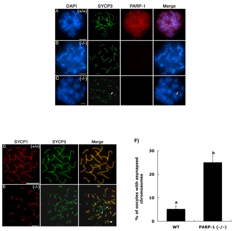
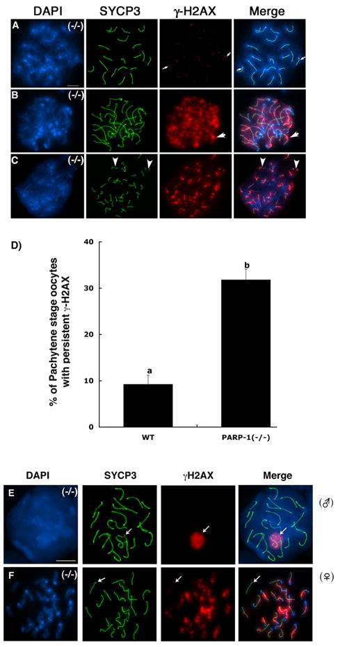
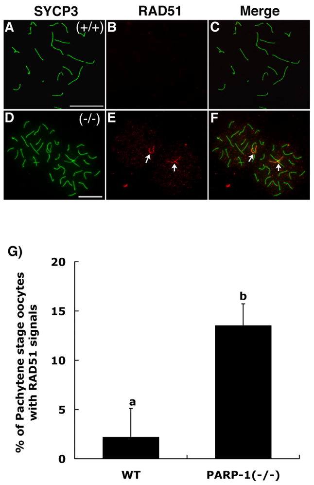
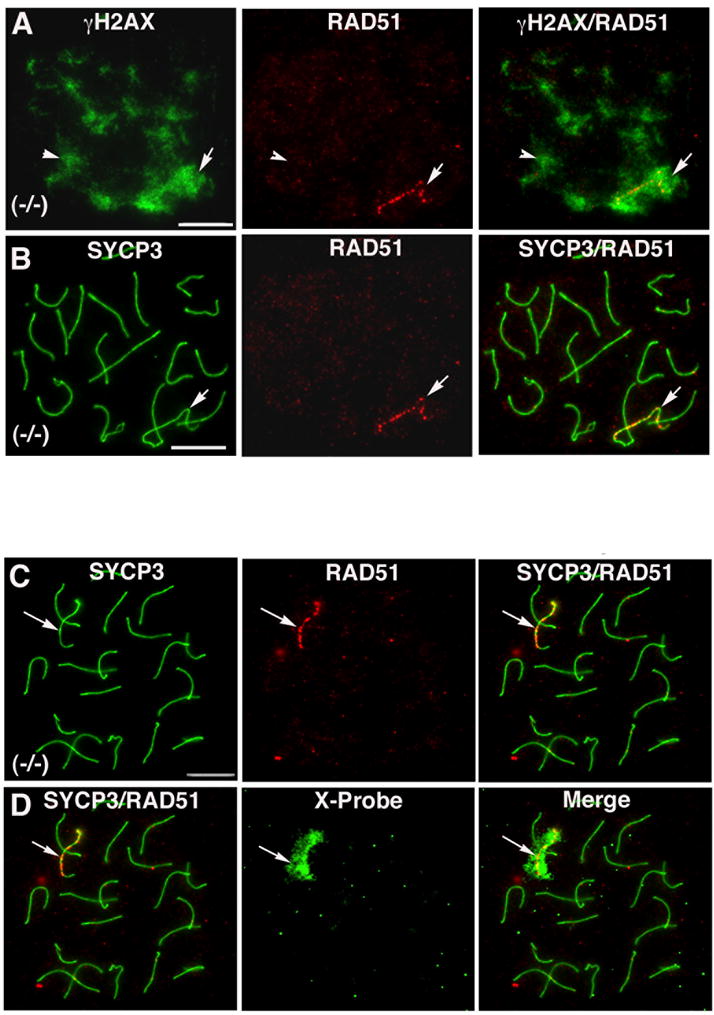
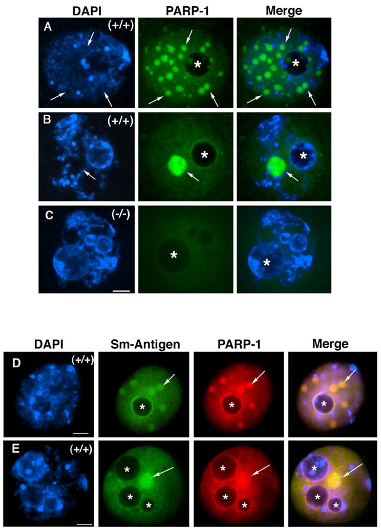
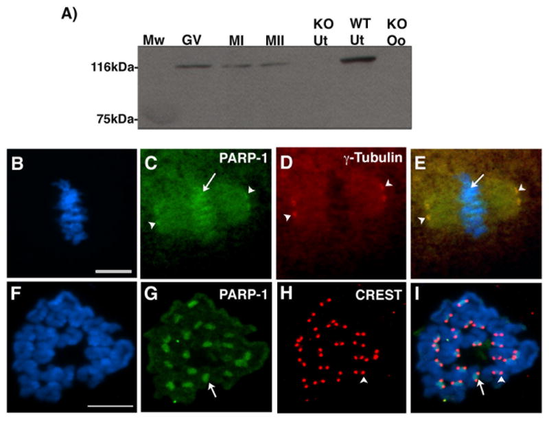
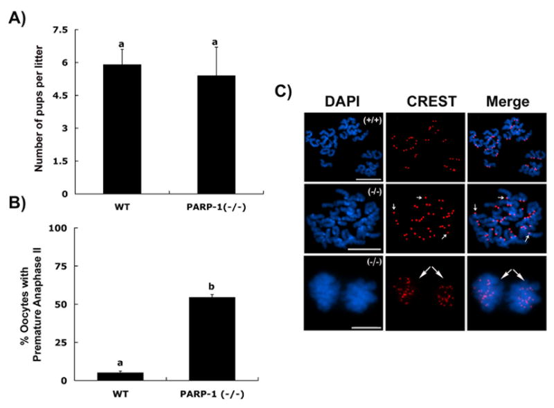
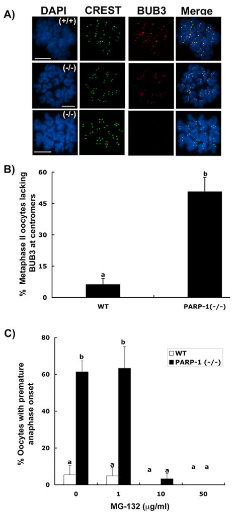
In spite of the impact of aneuploidy on human health little is known concerning the molecular mechanisms involved in the formation of structural or numerical chromosome abnormalities during meiosis. Here, we provide novel evidence indicating that lack of PARP-1 function during oogenesis predisposes the female gamete to genome instability. During prophase I of meiosis, a high proportion of Parp-1((-/-)) mouse oocytes exhibit a spectrum of meiotic defects including incomplete homologous chromosome synapsis or persistent histone H2AX phosphorylation in fully synapsed chromosomes at the late pachytene stage. Moreover, the X chromosome bivalent is also prone to exhibit persistent double strand DNA breaks (DSBs). In striking contrast, such defects were not detected in mutant pachytene spermatocytes. In fully-grown wild type oocytes at the germinal vesicle stage, PARP-1 protein associates with nuclear speckles and upon meiotic resumption, undergoes a striking re-localization towards spindle poles as well as pericentric heterochromatin domains at the metaphase II stage. Notably, a high proportion of in vivo matured Parp-1((-/-)) oocytes show lack of recruitment of the kinetochore-associated protein BUB3 to centromeric domains and fail to maintain metaphase II arrest. Defects in chromatin modifications in the form of persistent histone H2AX phosphorylation during prophase I of meiosis and deficient sister chromatid cohesion during metaphase II predispose mutant oocytes to premature anaphase II onset upon removal from the oviductal environment. Our results indicate that PARP-1 plays a critical role in the maintenance of chromosome stability at key stages of meiosis in the female germ line. Moreover, in the metaphase II stage oocyte PARP-1 is required for the regulation of centromere structure and function through a mechanism that involves the recruitment of BUB3 protein to centromeric domains.


Figures

Figure 1
Figure 1. Sub-cellular localization of the PARP-1 protein during prophase I of meiosis in mouse oocytes
(A) PARP-1 (red) exhibits a diffuse nuclear localization pattern in oocytes showing fully synapsed bivalents at the pachytene stage. The axial/lateral elements of the synaptonemal complex were stained with an antibody against the SYCP3 protein and are shown in green. (B) Pachytene stage oocyte obtained from Parp-1 null females showing fully synapsed bivalents lack detectable levels of PARP-1 protein, thus confirming antibody specificity. (C) A subpopulation of mutant oocytes exhibits a range of meiotic defects including incomplete homologous chromosome synapsis. (D) Co-localization of SYCP-1 and SYCP3 proteins at the pachytene stage confirms full synapsis of homologous chromosomes in wild-type oocytes. (E) The partial co-localization of SYCP1 and SYCP3 proteins observed in mutant oocytes revealed the extent of asynapsis between homologous chromosomes (arrows). (F) Analysis of meiotic configurations in wild type (WT) and Parp-1 (−/−) females revealed a significant increase (p<0.05) in the proportion of mutant oocytes that exhibit asynapsed bivalents. Scale bars=10 μm.
Figure 2
Figure 2. Persistence of γH2AX phosphorylation in late pachytene stage Parp-1 (−/−) oocytes
(A) Late pachytene stage oocyte obtained from a Parp-1 (−/−) female showing complete homologous chromosome synapsis (n=20 bivalents). At this stage, γH2AX phosphorylation is only detectable by the presence of a few small foci associated with synapsed bivalents. (B) A subpopulation of Parp-1 (−/−) oocytes at the late pachytene-early diplotene stage exhibits persistence of γH2AX phosphorylation throughout the chromatids of fully synapsed bivalents (arrows). (C) Abnormal meiosis in Parp-1 (−/−) oocytes showing intense γH2AX phosphorylation associated with each asynapsed chromosome and partial interruptions in SYCP3 staining suggesting the presence of chromosome breaks (arrowheads). (D) Proportion of mutant oocytes that exhibit persistent γH2AX phosphorylation in fully synapsed bivalents at the late pachytene-early diplotene stage. In contrast to the meiotic phenotype observed in the female germ line, pachytene stage spermatocytes (E) showed normal chromosome synapsis and γH2AX staining was found exclusively associated with the sex chromosome bivalent (arrow). Simultaneous staining of a mutant oocyte (F) reveals the extent of persistent γH2AX phosphorylation in the majority of chromosomes except for a single synapsed bivalent (arrow). Scale bars=10 μm.
Figure 3
Figure 3. Double strand DNA breaks (DSBs) persist at a single synapsed bivalent in Parp-1 (−/−) oocytes at the pachytene stage
(A) Wild type oocyte showing full synapsis of homologous chromosomes (20 bivalents) as determined by SYCP3 staining. (B) RAD51 foci (DSBs) are progressively resolved and hence become undetectable as meiotic chromosomes reach full synapsis in wild type oocytes. (C) Merge. (D–F) In contrast, persistence of RAD51 foci (red) at synapsed chromosomes (green) confirms the presence of unresolved DSBs at a single synapsed bivalent on each mutant oocyte (arrows). Note that two closely apposed pachytene stage oocytes are shown. (G) Proportion of wild type (WT) and mutant oocytes that exhibit persistent DSBs as labeled by RAD51 foci in a single bivalent (p<0.05). Scale bars=10 μm.
Figure 4
Figure 4. The X-chromosome bivalent fails to resolve DSBs in Parp-1 (−/−) oocytes
(A) A small proportion of mutant oocytes exhibit both persistent γH2AX phosphorylation (green) and RAD51 foci (red). However, in contrast with the wide distribution of γH2AX staining in the majority of synapsed chromosomes (arrowheads), RAD51 foci remain exclusively associated with a single chromosome bivalent (arrow). (B) Corresponding micrograph showing synapsed bivalents and localization of RAD51 foci in mutant oocytes. Immuno-FISH analysis revealed that in the absence of PARP-1 function, RAD51 foci (C; arrow) remain exclusively associated with the X-chromosome bivalent in mutant oocytes (D; arrow). Scale bars=10 μm.
Figure 5
Figure 5. PARP-1 associates with nuclear speckles in fully-grown oocytes at the GV stage
(A) Fully-grown wild type oocyte exhibiting decondensed chromatin typical of the non-surrounded nucleolus (NSN) configuration. In addition to its diffuse nuclear localization, PARP-1 forms several nuclear aggregates that accumulate at regions with no DAPI staining (arrows). The position of the nucleolus is indicated by (*). (B) Upon the transition into the surrounded nucleolus (SN) configuration, these nuclear aggregates coalesce into a prominent nuclear body that shows no association with DAPI-stained chromatin (arrows). (C) Parp-1 (−/−) oocyte showing only background fluorescence used as control for antibody specificity. (D–E) Representative images of wild type oocytes with the NSN configuration (D) and the SN configuration (E) showing diffuse nuclear staining as well as co-localization of PARP-1 protein with the Smith antigen (Sm), a marker for nuclear speckles. Note that PARP-1 can also be observed associated with the perinucleolar region. The position of the nucleolus is indicated by (*). Scale bars=10 μm.
Figure 6
Figure 6. PARP-1 associates with meiotic spindle poles and peri-centric heterochromatin in metaphase II stage oocytes
(A) Western blot analysis using wild type oocytes at the germinal vesicle (GV), metaphase I (MI) and metaphase II (MII) stage indicates that PARP-1 is a protein of approximately 116 kDA that shows no detectable change in electrophoretic mobility during oocyte meiotic maturation. Knockout uterus (KO Ut), knockout oocytes (KO Oo) and wild type uterus (WT Ut) were used as negative and positive controls, respectively. (B) Metaphase II stage chromosomes as shown by DAPI staining of whole mount oocytes. (C) PARP-1 is present at spindle poles (arrowheads) as well as the chromosomes of mature oocytes (arrows). (D) Corresponding micrograph showing the localization of γ-Tubulin signals at meiotic spindle poles. (E) Overlay. (F) High resolution chromosome spread at the metaphase II stage. (G) Association of PARP-1 with pericentric heterochromatin (arrow). Although PARP-1 signal can be detected at the chromatids of metaphase II stage chromosomes, the protein is highly enriched at pericentric domains. (H) Corresponding micrograph showing the localization of centromeres as detected by the CREST antiserum (arrowhead). (I) Overlay, note that the PARP-1 signal extends over a larger block of heterochromatin compared with the CREST signal. Scale bars=10 μm.
Figure 7
Figure 7. Evidence for a role of PARP-1 in sister chromatid cohesion in mouse oocytes
(A) Average litter size (±S.D.) in control and PARP-1 null females; (B) Percentage of oocytes showing premature anaphase-II onset upon removal from the oviductal environment. Different superscripts denote significant differences (p<0.05). (C) Top panel: Wild-type oocyte showing a normal chromosome complement with 40 CREST signals at the metaphase II stage. Middle panel: In vivo matured mutant oocyte showing the presence of several single chromatids (small arrows) at the metaphase II stage. Lower panel: Mutant oocyte showing premature anaphase-II onset ex vivo following exposure to the culture environment for 15 minutes. Long arrows indicate the premature segregation of sister chromatids (20 CREST signals each) into opposite poles. Scale bars=10 μm.
Figure 8
Figure 8. Parp-1 (−/−) oocytes fail to recruit BUB3 protein to the centromere at the metaphase-II stage
(A) Top panel: Specific co-localization of the CREST signals (green) with the BUB3 protein (red) at the centromeres of wild-type metaphase-II oocytes. Center panel: Similar to wild-type oocytes, some mutant oocytes show correct localization of the BUB3 protein at centromeres. Lower panel: A high proportion of Parp-1(−/−) oocytes fail to recruit BUB3 to meiotic centromeres, yet show proper CREST staining. Scale bars=10 μm. (B) Proportion of metaphase-II oocytes lacking BUB3 protein at centromeres; data are presented as the mean (±S.D.) after three independent experiments (p<0.05). (C) Proportion of wild type (WT) and mutant oocytes that exhibit premature anaphase onset in the presence or absence of the proteasome inhibitor MG-132 (p<0.05).

Similar articles

Cited by

References

    1. Ahel I, Ahel D, Matsusaka T, Clark AJ, Pines J, Boulton SJ, West SC. Poly(ADP-ribose)-binding zinc finger motifs in DNA repair/checkpoint proteins. Nature. 2008;451:81–5. - PubMed
    1. Althaus FRHL, Kleczkowska HE, Malanga M, Naegeli H, Panzeter PL, Realini CA. Histone shuttling by poly ADP-ribosylation. Mol Cell Biochem. 1994;138:53–9. - PubMed
    1. Ame JC, Spelenhauer C, de Murcia G. The PARP superfamily. Bioessays. 2004;26:882–93. - PubMed
    1. Baker SM, Plug AW, Prolla TA, Bronner CE, Harris AC, Yao X, Christie DM, Monell C, Arnheim N, Bradley A, Ashley T, Liskay RM. Involvement of mouse Mlh1 in DNA mismatch repair and meiotic crossing over. Nat Genet. 1996;13:336–42. - PubMed
    1. Bakkenist CJ, Kastan MB. DNA damage activates ATM through intermolecular autophosphorylation and dimer dissociation. Nature. 2003;421:499–506. - PubMed

Publication types

LinkOut - more resources