Review
. 2009 Oct;1790(10):1095-108.
doi: 10.1016/j.bbagen.2009.05.008. Epub 2009 May 20.

Lens aging: effects of crystallins

Affiliations
Review

Lens aging: effects of crystallins

K Krishna Sharma et al. Biochim Biophys Acta. 2009 Oct.

Abstract

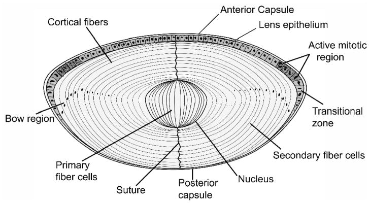
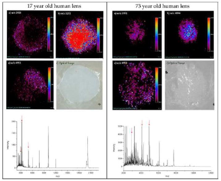
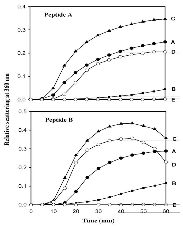
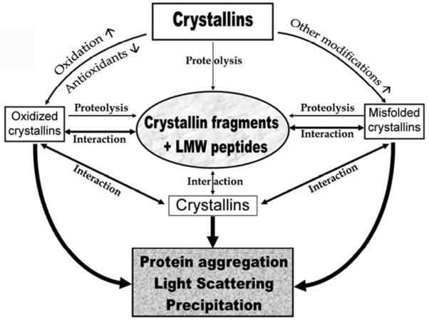
The primary function of the eye lens is to focus light on the retina. The major proteins in the lens--alpha, beta, and gamma-crystallins--are constantly subjected to age-related changes such as oxidation, deamidation, truncation, glycation, and methylation. Such age-related modifications are cumulative and affect crystallin structure and function. With time, the modified crystallins aggregate, causing the lens to increasingly scatter light on the retina instead of focusing light on it and causing the lens to lose its transparency gradually and become opaque. Age-related lens opacity, or cataract, is the major cause of blindness worldwide. We review deamidation, and glycation that occur in the lenses during aging keeping in mind the structural and functional changes that these modifications bring about in the proteins. In addition, we review proteolysis and discuss recent observations on how crystallin fragments generated in vivo, through their anti-chaperone activity may cause crystallin aggregation in aging lenses. We also review hyperbaric oxygen treatment induced guinea pig and 'humanized' ascorbate transporting mouse models as suitable options for studies on age-related changes in lens proteins.


Figures

Fig. 1
Fig. 1. A drawing of lens showing the single layer of epithelial cells covering the anterior cross section
The elongated fiber cells are directly in contact with epithelial layer in the anterior region, whereas they make contact with capsule in the posterior region. In the lens bow region the cells differentiate, elongate, lose their organelles and begin to form newly differentiated fiber cells.
Fig. 2
Fig. 2. Mass distribution in the water-soluble fraction of young (19 years, solid line and filled circle) and aged (83 years, broken line and open circle) human lenses using multi-angle light scattering
The samples (250 mg of proteins) were injected into a TSK G5000PWXL size exclusion column attached to a Shimadzu high pressure liquid chromatograph system with refractive index detector. The eluting proteins were analyzed with the help of Wyatt multi-angle light scattering and quasi-elastic light scattering detectors. The mobile phase contained 0.05 M sodium phosphate and 0.15 m sodium chloride, pH 7.2. The mass was determined using ASTRA software from Wyatt. Reproduced with permission from the J. of Biol. Chem. (2008) 283 8477–8485.
Fig. 3
Fig. 3. Schematic representation of the modification of aB-crystallin-K90C to K90C-AE [aminoethylation] and K90C-OP [3-oxidopyridinium ethylation]
Reproduced with permission from Biochemistry (2008) 46, 14682–92.
Fig. 4
Fig. 4. Aggregation assay with wild-type aB-crystallin, K90C, K90C-AE, and K90C-OP using alcohol dehydrogenase [ADH] as client protein
The assays were carried out as described earlier [120] using 1:10 w/w ratio of aB-crystallin (native or modified) and ADH. Reproduced with permission from Biochemistry (2008) 46, 14682–92.
Fig. 5
Fig. 5. MALDIimaging of a 17- and a 73-year-old human lens
The three ion images correspond to three distinct molecular species measured from a 12 micron section. The three ion images shown in 17-year-old human lens have m/z values of 2928, 3252, and 4911. The three ion images shown in 73-year-old human lens have m/z values of 2931, 4096, and 4911. The optical image [d] of the tissue specimen shown was captured following application of sinapinic acid matrix by spray coating. The data were compiled into Analyze 7.5 format after baseline correction. A 2-D ion density map (image) for specific ion constructed is shown. The procedure allows the construction of images for the specific mass signal detected from the surface of the section, theoretically offering an opportunity to generate hundreds of images, each representing the distribution of the specific ionized species from a single imaging mass spectrometry experiment. Average mass spectrum collected from the surface of the lens samples are shown in the bottom of the figure. Notice the abundance of ions in the spectrum for 73-year-old human lens. Red arrows denote the ions highlighted in figures. Data in this Figure is unpublished results from authors study.
Fig. 6
Fig. 6. Inhibition of chaperone-like activity of a-crystallin against bL-crystallin by peptide A and B
Peptide A: aB-1MDIAIHHPWRRPFFPFH18; Peptide B: bA3/A159SNAYHIERLMSFRPIC74Top, anti-chaperone activity of Peptide A. Bottom, anti-chaperone activity of peptide B. A, 100 mg of bL-crystallin; B, bL-crystallin + 25 mg of aA-crystallin; C, bL-crystallin + 60 mg of peptide; D, bL-crystallin + aA-crystallin + peptide; E, peptide alone (60 mg). The aggregation assay was carried out as described earlier [185]. Reproduced with permission from the Journal of Biol. Chem. (2008) 283 8477–8485.
Figure 7
Figure 7. Schematic representation of the proposed role of crystallin fragments in lenticular aging and cataractogenesis
Oxidizing and modifying reactions/factors are not shown for simplicity.

Similar articles

Cited by

References

    1. Dilley KJ, Pirie A. Changes to the proteins of the human lens nucleus in cataract. Exp Eye Res. 1974;19:59–72. - PubMed
    1. Spector A, Li S, Sigelman J. Age-dependent changes in the molecular size of human lens proteins and their relationship to light scatter. Invest Ophthalmol. 1974;13:795–798. - PubMed
    1. Roy D, Spector A. Absence of low-molecular-weight alpha crystallin in nuclear region of old human lenses. Proc Natl Acad Sci USA. 1976;73:3484–3487. - PMC - PubMed
    1. Bloemendal H. The lens proteins. In: Bloemendal H, editor. Molecular and Cellular Biology of the Eye Lens. New York: John Willey & Sons; 1981. pp. 1–49.
    1. Jaffe NS, Horwitz J. Lens and Cataract. In: Podos SM, Yanoff M, editors. Text Book of Ophthalmology. Vol. 3. Gower Med. Publishing; New York: 1991.

Publication types