. 2009 Sep;42(1):33-44.
doi: 10.1016/j.mcn.2009.05.003. Epub 2009 May 20.

Fast inactivation of Shal (K(v)4) K+ channels is regulated by the novel interactor SKIP3 in Drosophila neurons

Affiliations

Fast inactivation of Shal (K(v)4) K+ channels is regulated by the novel interactor SKIP3 in Drosophila neurons

Fengqiu Diao et al. Mol Cell Neurosci. 2009 Sep.

Abstract

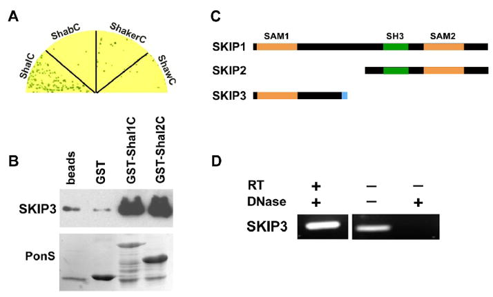
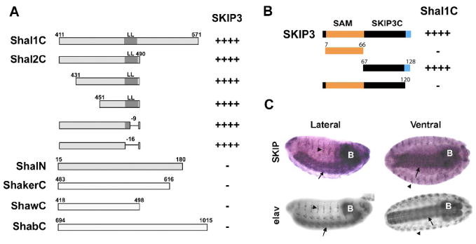
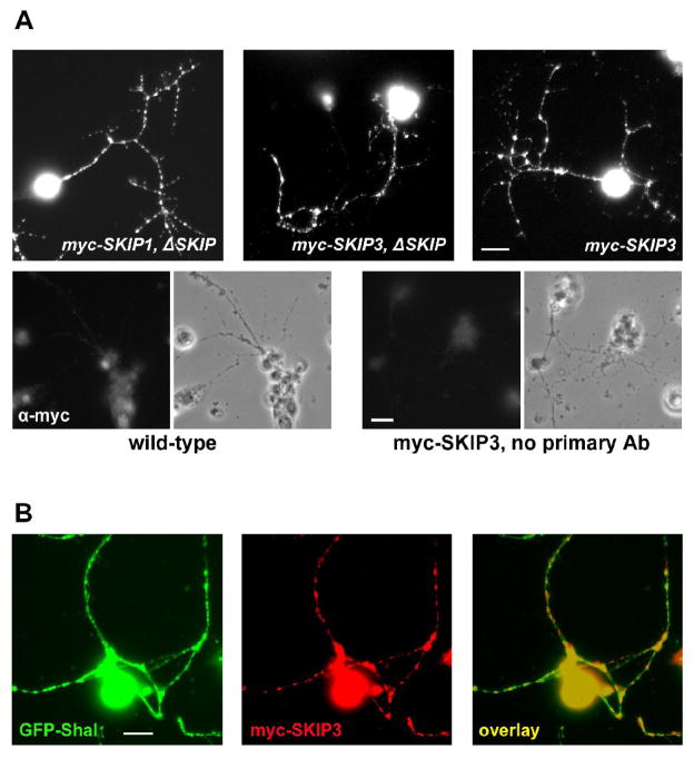
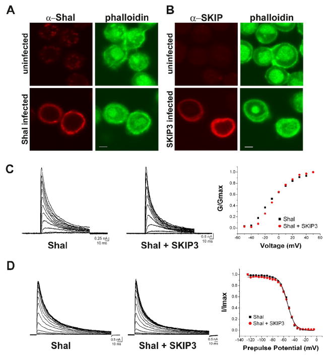
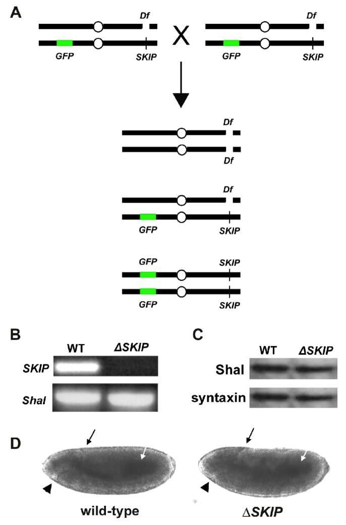
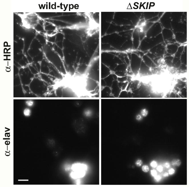
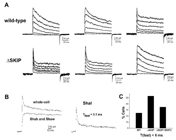
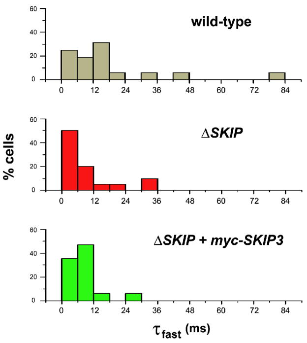
Shal K+ (K(v)4) channels across species carry the major A-type K+ current present in neurons. Shal currents are activated by small EPSPs and modulate post-synaptic potentials, backpropagation of action potentials, and induction of LTP. Fast inactivation of Shal channels regulates the impact of this post-synaptic modulation. Here, we introduce SKIP3, as the first protein interactor of Drosophila Shal K+ channels. The SKIP gene encodes three isoforms with multiple protein-protein interaction domains. SKIP3 is nervous system specific and co-localizes with Shal channels in neuronal cell bodies, and in puncta along processes. Using a genetic deficiency of SKIP, we show that the proportion of neurons displaying a very fast inactivation, consistent with Shal channels exclusively in a "fast" gating mode, is increased in the absence of SKIP3. As a scaffold-like protein, SKIP3 is likely to lead to the identification of a novel regulatory complex that modulates Shal channel inactivation.


Figures

Fig. 1
Fig. 1. The Novel Protein, SKIP3, Interacts with the C-terminus of Shal K+ Channels
(A) SKIP3 was tested in direct Y2H tests with the C-termini of representative isoforms of voltage-gated K+ channels from the four major subfamilies, including Shal1 (ShalC), ShA1 (ShakerC), Shab-RB (ShabC), and Shaw2 (ShawC). Shown are co-transfected yeast grown on stringent media and X-α-Gal to select for interaction. Note that growth and a blue color, indicating interaction, is specific for ShalC. (B) Shown are results from GST-pull down assays using GST-fused to the C-termini of Shal1 (residues 411 to 571) and Shal2 (residues 411 to 490), then incubated with purified SKIP3 protein; glutathione-agarose beads alone (beads) and GST were used as negative controls. A representative immunoblot of the proteins pulled-down by GST-fusion proteins (top) shows that large amounts of SKIP3 are pulled-down by GST-Shal1C and GST-Shal2C, with much lower background levels pulled-down by beads alone or GST. Immunoblot was incubated with Ponceau S (PonsS) to confirm that GST protein(s) were indeed pulled-down by glutathione-beads. (C) SKIP encodes three alternatively spliced isoforms, SKIP1 (CG31163-PB/PC), SKIP2 (CG31163-PA), and SKIP3. Each isoform contains a different combination of protein binding domains, including two different SAM domains (orange) and an SH3 domain (green) domain. SKIP3 contains a unique eight residue C-terminal sequence (blue), which is required for binding Shal channels (see Fig. 2). (D) RT-PCR of SKIP3 from wild-type embryos. When RNA treated with DNase was used as template, SKIP3 was successfully reverse-transcribed and amplified by PCR (RT-PCR, left lane), suggesting that SKIP3 is a true isoform expressed in the embryo. When RNA was not treated with DNase, as indicated (−), SKIP3 could be amplified even without reverse transcription (middle lane), suggesting that DNase treatment is essential to degrade contaminating genomic DNA. A mock RT-PCR performed from DNase-treated RNA in the absence of reverse transcriptase (RT) confirmed that SKIP3 was not amplified without RT.
Fig. 2
Fig. 2. Mapping the SKIP3-Shal Interaction, and Nervous System Specific Expression of SKIP3
(A) Direct Y2H results are shown for “bait” fusion proteins, including C-terminal sequences from Shal1 (Shal1C), Shal2 (Shal2C), ShA1 (ShakerC), Shab-RB (ShabC), and Shaw2 (ShawC), and the N-terminus of Shal (ShalN), co-expressed with the SKIP3 “prey” fusion protein identified in our Y2H screen. Positive interactions, assayed by growth on stringent media and a blue color when grown on X-α-gal, are indicated by the number of “+” symbols; “−” indicates no/minimal growth or blue color. SKIP3 interacted specifically with the C-terminus of Shal. Binding is mediated by the sequence between residues 451 and 474 since deletions outside of this region, including the conserved di-leucine motif (LL), did not affect the interaction. (B) Direct Y2H results are shown for the Shal1C “bait” co-expressed with various truncations of SKIP3 as “prey”. Full-length SKIP3, containing a 60 amino acid N-terminal SAM domain (orange), followed by a 54 amino acid domain (SKIP3C; black), and a C-terminal eight amino acids (blue) unique to SKIP3, showed strong interaction with Shal1C, as expected. Protein-protein interaction, indicated by the number of “+” symbols, was assayed by growth on stringent media and a blue color when grown on X-α-Gal. Note that truncation of the C-terminal eight amino acids (blue) of SKIP3 completely disrupted interaction with Shal1C, while deletion of the SAM domain did not. Similar results were seen for Shal2C (data not shown). (C) Representative RNA in situ hybridization for SKIP (top) and immunostaining for the neuron-specific elav protein (bottom) in embryos 12 hours AEL. The single-stranded digioxygenin-labeled probe for in situs, amplified from SKIP3 template, likely hybridizes with both SKIP1 and SKIP3. After hybridization, the embryos were incubated with anti-Dig antibody and developed by NBT/BCIP. SKIP1/3 displays expression primarily in the central nervous system (brain indicated by “B”, ventral nerve cord indicated by arrows), and peripheral nervous system (indicated by arrowheads), similar to elav.
Fig. 3
Fig. 3. Subcellular Localization of myc-SKIP1, myc-SKIP3, and GFP-Shal in Embryonic Neurons
(A) Transgenic lines were generated expressing the neuron-specific elav-GAL4 driver and either the transgene UAS-myc-SKIP1 or UAS-myc-SKIP3 (noted as elav:myc-SKIP1 or elav:myc-SKIP3). Additionally, elav:myc-SKIP1 was present either in a wild-type background or a homozygous Df(3R)Exel6190SKIP) background (see text and Fig. 5). Primary cultures were made from embryos of these genotypes and immunostained for myc. Shown are representative single neurons expressing myc-SKIP1/3 in a ΔSKIP background (myc-SKIP1/3, ΔSKIP) and myc-SKIP3 in a wild-type background (myc-SKIP3). myc-SKIP1 and myc-SKIP3 are localized in cell bodies and in puncta along neuronal processes. As negative controls, clusters of wild-type neurons were immunostained with the anti-myc antibody neurons (bottom, left) and myc-SKIP3 expressing clusters of neurons were stained with only secondary antibody (bottom, right); phase-contrast images are shown to the right of each stained panel. (B) Co-localization of myc-SKIP3 and GFP-Shal was examined in cultures from transgenic lines in which elav-GAL4 is driving expression of UAS-myc-SKIP3 and UAS-GFP-Shal2. Shown is a representative image of neurons immunostained for myc-SKIP3 (red) and GFP-Shal (green). Co-localization (yellow) of myc-SKIP3 and GFP-Shal is observed in cell bodies, and in many, although not all, puncta in neuronal processes (right). All scale bars represent 5 μm.
Fig. 4
Fig. 4. Co-Expression of Shal and SKIP3 in Sf9 Cells
(A–B) Sf9 cells were infected with baculovirus expressing either Shal2 (A) or SKIP3 (B). Representative confocal images of Sf9 cells immunostained for Shal/SKIP3, and FITC-conjugated phalloidin, show expression of Shal and SKIP3. Uninfected cells are shown for comparison. Scale bars represent 5 μm. (C) Whole-cell voltage-clamp recordings from representative Sf9 cells infected with the Shal-expressing baculovirus alone (Shal), or both Shal-expressing and SKIP3-expressing baculoviruses (Shal + SKIP3); capacitative transients have been clipped for illustration purposes. Left and middle, Current traces in response to 120 ms voltage-jumps were taken from a holding potential of −100 mV to voltages between −50 mV and +50 mV, in 10 mV increments. Right, G/Gmax-Voltage plots are shown for each cell; no significant difference was observed in any cells. (D) Left and middle, Representative current traces in response to a 140 ms test potential to +50 mV, following a 500 ms pre-pulse at potentials from −125 to −40 mV, in 5 mV increments. Right, Resulting steady-state inactivation plots and fits are shown from these two cells. No significant differences were observed in any cells. Also see Table 1.
Fig. 5
Fig. 5. Genetic Deficiency Df(3R)Exel6190 of SKIP as a Tool to Study the Role of SKIP
(A) Shown is the genetic strategy used to identify embryos and embryonic cultures that are homozygous for the deficiency Df(3R)Exel6190 (Df). Since the Df(3R)Exel6190 deficiency is homozygous lethal in adult flies, heterozygotes are maintained over a balancer chromosome containing a transgene for GFP expression. Heterozygous adult flies crossed together produce viable embryos of the three genotypes shown. Homozygous Df(3R)Exel6190 embryos (and primary embryonic cultures) can be identified by the lack of GFP expression. (B) PCR amplifications of SKIP and Shal from genomic DNA isolated from wild-type and homozygous Df(3R)Exel6190SKIP) embryos. Df(3R)Exel6190 was maintained as a heterozygote over a GFP balancer chromosome. Heterozygotes were crossed to each other, embryos were collected 13–14 hours AEL, and homozygous Df(3R)Exel6190 embryos were selected based on the absence of GFP expression (ΔSKIP). GFP-positive embryos, containing at least one copy of the genes uncovered by the deficiency, were used as a wild-type control (WT). PCR using primers for a common SKIP sequence and Shal show that SKIP could not be amplified from homozygous Df(3R)Exel6190 embryos, verifying that Df(3R)Exel6190 does indeed remove the SKIP gene. (C) Representative immunoblot analysis for Shal protein in wild-type (WT) and homozygous SKIP deficiency (ΔSKIP) embryos. Embryos 13–14 hours AEL were identified as in (B), and analyzed by immunoblot analysis (20 embryos/lane), using antibodies against Shal and syntaxin. Levels of Shal protein are similar in wild-type and ΔSKIP embryos. (D) Representative wild-type and homozygous Df(3R)Exel6190 embryos at stage 10 (5–6 hours AEL), at which time cells are dissociated to make primary cultures, are shown. No gross differences can be observed in the developing embryos at this stage. Black arrows indicate the dorsal invagination of the posterior midgut primordium, white arrows indicate the midgut primordium, and black arrowheads indicate the stomodium.
Fig. 6
Fig. 6. Primary Cultures from Df(3R)Exel6190 Embryos are Similar to Wild-Type
Cells were dissociated from wild-type and homozygous Df(3R)Exel6190SKIP) embryos, and grown for 2–5 days in culture. Cells were fixed and immunostained using α-elav and α-horseradish peroxidase (α-HRP) antibodies, which recognize a neuron-specific RNA binding protein and all neuronal membranes, respectively. Representative images of small clusters of neurons from these cultures are shown. The number of neuronal cell bodies recognized by α-elav and extent of neuronal processes recognized by α-HRP were indistinguishable from one another across many cultures.
Fig. 7
Fig. 7. The Inactivation Rate of Shal K+ Currents is Affected by the Loss of SKIP3
(A) Whole-cell voltage-clamp recordings of three representative wild-type (top row) and Df(3R)Exel6190 (ΔSKIP, bottom row) neurons are shown. K+ currents were activated by 120 ms voltage jumps to −50 to +50 mV, in 10 mV increments, from a holding potential of −100 mV; capacitative transients have been clipped for illustration purposes. Inactivation rates varied from cell to cell, however, almost twice as many Df(3R)Exel6190 neurons displayed the very fast inactivating A-type currents (bottom row, left and middle recordings). (B) Separation of Shal current from Shab/Shaw currents by pre-pulse potential. In one representative neuron, the whole-cell K+ current is shown in response to a voltage jump to +50 mV from a 500 ms pre-pulse of −125 mV (left, top trace). With a pre-pulse of −45 mV, the transient A-type current encoded by Shal is completely inactivated and only the delayed-rectifier currents encoded by Shab and Shaw are activated (left, bottom trace). Subtraction of the Shab/Shaw currents from the whole-cell current yields the Shal K+ current (right). In this neuron, the inactivation time course of the Shal current (first 30 ms after peak) is fit with a single exponential function (best fit with τfast =3.1 ms). (C) Quantification of the percentage of wild-type (WT) and Df(3R)Exel6190 (ΔSKIP) neurons containing a Shal current with an inactivation rate that corresponds to Shal channels exclusively in the “fast” gating mode (τfast< 6 ms). Shal currents were isolated as in (B), and the inactivation time course 30 ms following peak current was fit with the single exponential decay function, y(t)=Ae(−t/τfast) + c. In contrast to 25% of wild-type neurons (N=16), 55% of Df(3R)Exel6190 (ΔSKIP) neurons (N=20) contained a Shal current with τfast< 6 ms. Transgenic expression of myc-SKIP3 in Df(3R)Exel6190 neurons (ΔSKIP + SKIP3) showed partial rescue, with 35% (N=17) containing a Shal current with τfast< 6 ms.
Fig. 8
Fig. 8. Distribution of Fast Inactivation Rates of Shal K+ Currents in Wild-Type and ΔSKIP Neurons
Histograms of fast inactivation time constants (τfast) from whole-cell Shal currents isolated from wild-type (top), Df(3R)Exel6190 (middle; ΔSKIP), and transgenic myc-SKIP3 in Df(3R)Exel6190 (bottom; ΔSKIP + myc-SKIP3) neurons. Shal K+ currents elicited with a voltage jump to +50 mV were isolated as described in Fig. 7B. Inactivation time courses 30 ms following peak current were fit with the single exponential decay function, y(t)=Ae(−t/τfast) + c. X-axis represents τfast values binned every 6 ms; y-axis represents the % cells (N=16 for wild-type, N=20 for ΔSKIP, N=17 for ΔSKIP + myc-SKIP3). Note the shift in τfast values to faster inactivation rates in SKIP neurons compared with wild-type.

Similar articles

Cited by

References

    1. Alzheimer C, Schwindt PC, Crill WE. Modal gating of Na+ channels as a mechanism of persistent Na+ current in pyramidal neurons from rat and cat sensorimotor cortex. J Neurosci. 1993;13:660–673. - PMC - PubMed
    1. An WF, Bowlby MR, Betty M, Cao J, Ling HP, Mendoza G, Hinson JW, Mattsson KI, Strassle BW, Trimmer JS, Rhodes KJ. Modulation of A-type potassium channels by a family of calcium sensors. Nature. 2000;403:553–556. - PubMed
    1. Bhattacharjya S, Xu P, Chakrapani M, Johnston L, Ni F. Polymerization of the SAM domain of MAPKKK Ste11 from the budding yeast: implications for efficient signaling through the MAPK cascades. Protein Sci. 2005;14:828–835. - PMC - PubMed
    1. Brand A, Perrimon N. Targeted gene expression as a means of altering cell fates and generating dominant phenotypes. Development. 1993;118:401–415. - PubMed
    1. Brock MW, Lebaric ZN, Neumeister H, Detomaso A, Gilly WF. Temperature-dependent expression of a squid Kv1 channel in Sf9 cells and functional comparison with the native delayed rectifier. J Membr Biol. 2001;180:147–161. - PubMed

Publication types

MeSH terms

Substances