. 2009 Jul 20;390(1):1-12.
doi: 10.1016/j.virol.2009.04.011. Epub 2009 May 22.

Pathological manifestations of feline immunodeficiency virus (FIV) infection in wild African lions

Affiliations

Pathological manifestations of feline immunodeficiency virus (FIV) infection in wild African lions

Melody E Roelke et al. Virology. .

Abstract

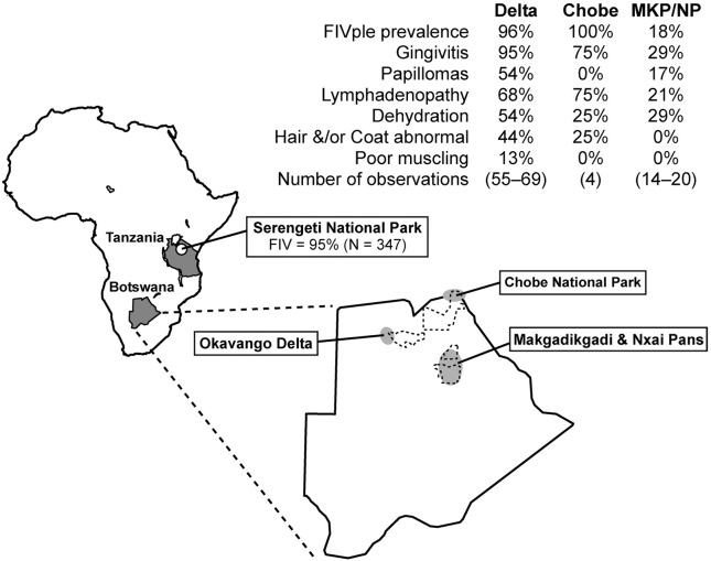
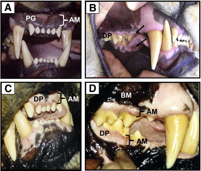
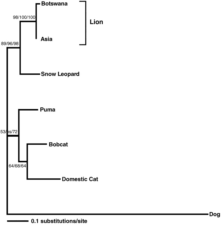
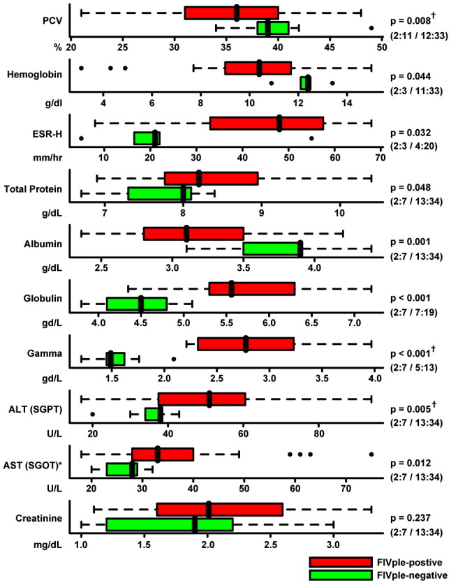
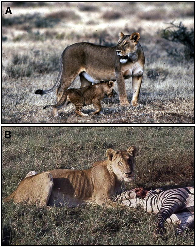
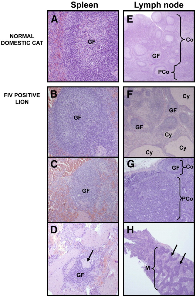
Feline immunodeficiency virus (FIV) causes AIDS in the domestic cat (Felis catus) but has not been explicitly associated with AIDS pathology in any of the eight free-ranging species of Felidae that are endemic with circulating FIV strains. African lion (Panthera leo) populations are infected with lion-specific FIV strains (FIVple), yet there remains uncertainty about the degree to which FIV infection impacts their health. Reported CD4+ T-lymphocyte depletion in FIVple-infected lions and anecdotal reports of lion morbidity associated with FIV seroprevalence emphasize the concern as to whether FIVple is innocuous or pathogenic. Here we monitored clinical, biochemical, histological and serological parameters among FIVple-positive (N=47) as compared to FIVple-negative (N=17) lions anesthetized and sampled on multiple occasions between 1999 and 2006 in Botswana. Relative to uninfected lions, FIVple-infected lions displayed a significant elevation in the prevalence of AIDS-defining conditions: lymphadenopathy, gingivitis, tongue papillomas, dehydration, and poor coat condition, as well as displaying abnormal red blood cell parameters, depressed serum albumin, and elevated liver enzymes and gamma globulin. Spleen and lymph node biopsies from free-ranging FIVple-infected lions (N=9) revealed evidence of lymphoid depletion, the hallmark pathology documented in immunodeficiency virus infections of humans (HIV-1), macaques, and domestic cats. We conclude that over time FIVple infections in free-ranging lions can lead to adverse clinical, immunological, and pathological outcomes in some individuals that parallel sequelae caused by lentivirus infection in humans (HIV), Asian macaques (SIV) and domestic cats (FIVfca).


Figures

Fig. 1
Fig. 1
Study sites in Botswana and Tanzania. Dotted outlines designate national parkland, while shaded ovals encompass the general areas where lions were sampled. N indicates the number of lions tested for FIV. Not all parameters were determined for every animal handled at every time point, therefore N was not the same for all clinical observations from a region and is given as a range.
Fig. 2
Fig. 2
Gingival tissue of normal (A) and FIV-positive lions with varying stages of gingivitis (B–D); frontal incisors (A and C) and lateral molar (B and D) views. (A) Normal, healthy gingiva in an FIVple-negative lion. Note darkly pigmented (PG) alveolar mucosa (AM) (adhered to bone). (B) Early stage FIVple gingivitis in a young adult female lion showing linear gingival erythema (arrow), an inflammatory process that starts to erode the normal darkly pigmented alveolar mucosa resulting in a narrow zone of depigmentation (DP) along free edge of gingiva adjacent to molars and lower outside incisors. (C and D) Granulomatous gingivitis typically seen in all mature, chronically FIV-infected lions. Note extensive depigmented zone affecting virtually all alveolar gingival mucosa (AM) but sparing the bucal mucosa (BM). The depigmented area above the upper canine teeth is normal.
Fig. 3
Fig. 3
Phylogeny of concatenated early and late genes (900 bp) of a Botswana lion papilloma virus compared to papilloma viruses isolated from several felid species. Canine papilloma virus was used as an outgroup. Shown here is the maximum likelihood (ML) tree. Minimum evolution (ME) and maximum parsimony (MP) trees gave similar topologies. Bootstrap values are given at all nodes (ML/ME/MP; ns = not significant).
Fig. 4
Fig. 4
Comparison of blood parameters, protein levels, and chemistry values from FIV-positive (red) and negative (green) lions. The black vertical line represents the median, colored bars represent the interquartile range (IQR), and dotted lines represent the range up to 1.5 times the IQR. Outliers are represented by open circles, and biomarkers with an asterisk (ALT and AST) had extreme outliers that are not shown here in the interest of scale. Due to the non-normal distribution of most biomarkers, non-parametric Wilcoxon rank-sum statistics were used to determine p values. Number of FIVple-negative and positive male and female lions is indicated in parentheses (negative males:negative females/positive males:positive females). Among FIVple-positive lions, all of the parameters in this figure were examined for age-dependant effects. The three traits marked with a “†” had a significant difference in values between adult (4–8 years) and older (9+) age classes, with older lions trending further away from the mean of the FIVple-negative lions. However, when older lions were removed from the analysis of these three parameters, significant differences still remained between FIVple-positive and -negative lions (p = 0.029, p = 0.0001, and p = 0.032 for PCV, globulin, and ALT, respectively). Abbreviations: PCV — packed cell volume; ESR — erythrocyte sedimentation rate; ALT — alanine aminotransferase (aka SGPT); AST — aspartate aminotransferase (aka SGOT).
Fig. 5
Fig. 5
Varying degrees of body condition in FIVple-infected animals. While most uninfected and infected lions appear to be in good to excellent physical condition (A; 94-184 Ple July 1994, Serengeti National Park; photo credit Christopher Ratier). However, occasionally lions with unexplained weight loss (cachexia) are observed in populations with FIVple-positive lions (B; 94-060 Ple Mar 1994, Serengeti National Park; similar “poor doers” have been seen in Kruger National Park and the Okavango Delta; photo credit Melody Roelke), often in the context of secondary disease outbreaks. Both these lions were photographed in the spring of 1994 during a CDV epidemic. CDV alone is not commonly associated with wasting.
Fig. 6
Fig. 6
Spleen (A–D) and lymph node (E–H) tissue from normal, uninfected domestic cats (A and E) and FIVple-infected lions (B–D and F–H). GF = germinal follicle; Co = cortex; PCo = paracortex; Cy = cyst; M = medulla. (A) Healthy splenic tissue. (B) A hyperplastic follicle. (C) Follicular atrophy. (D) Severe follicular atrophy with pronounced lymphoid depletion. Arrow indicates an area of hyaline depletion. (E) A healthy lymph node with many follicles in the cortex and a smaller paracortical area. (F) Mixed hyperplastic follicles with cystic areas. (G) Cortical atrophy with paracortical hyperplasia. (H) Extensive medullary plasmacytosis. Arrows indicate cords of plasma cells.

Similar articles

Cited by

References

    1. R Development Core Team, 2008. R: A language and environment for statistical computing. R Foundation for Statistical Computing, Vienna, Austria.
    1. Ackley C.D., Yamamoto J.K., Levy N., Pedersen N.C., Cooper M.D. Immunologic abnormalities in pathogen-free cats experimentally infected with feline immunodeficiency virus. J. Virol. 1990;64(11):5652–5655. - PMC - PubMed
    1. Antunes A., Troyer J.L., Roelke M.E., Pecon-Slattery J., Packer C., Winterbach C., Winterbach H., Hemson G., Frank L., Stander P., Siefert L., Driciru M., Funston P.J., Alexander K.A., Prager K.C., Mills G., Wildt D., Bush M., O'Brien S.J., Johnson W.E. The evolutionary dynamics of lion Panthera leo revealed by host and viral population genomics. PLOS Genetics. 2008;4(11):e1000251. - PMC - PubMed
    1. Barlough J.E., Ackley C.D., George J.W., Levy N., Acevedo R., Moore P.F., Rideout B.A., Cooper M.D., Pedersen N.C. Acquired immune dysfunction in cats with experimentally induced feline immunodeficiency virus infection: comparison of short-term and long-term infections. J. Acquir. Immune Defic. Syndr. 1991;4(3):219–227. - PubMed
    1. Bendinelli M., Pistello M., Lombardi S., Poli A., Garzelli C., Matteucci D., Ceccherini-Nelli L., Malvaldi G., Tozzini F. Feline immunodeficiency virus: an interesting model for AIDS studies and an important cat pathogen. Clin. Microbiol. Rev. 1995;8(1):87–112. - PMC - PubMed

Publication types

MeSH terms