

Assessing functional outcomes following intracerebral hemorrhage in rats
- PMID: 19464275
- PMCID: PMC6918942
- DOI: 10.1016/j.brainres.2009.05.038
Assessing functional outcomes following intracerebral hemorrhage in rats
Abstract
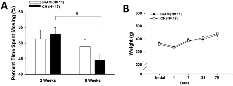
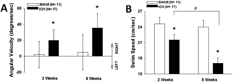
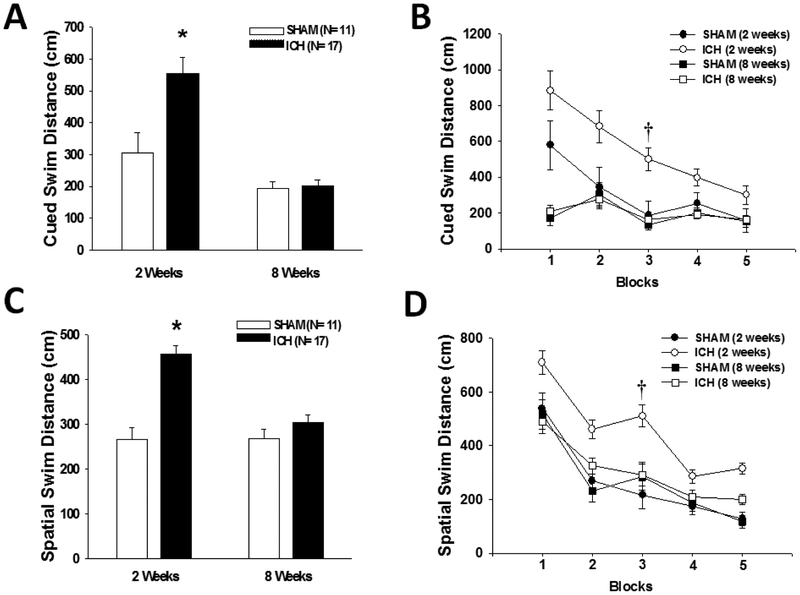
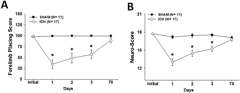
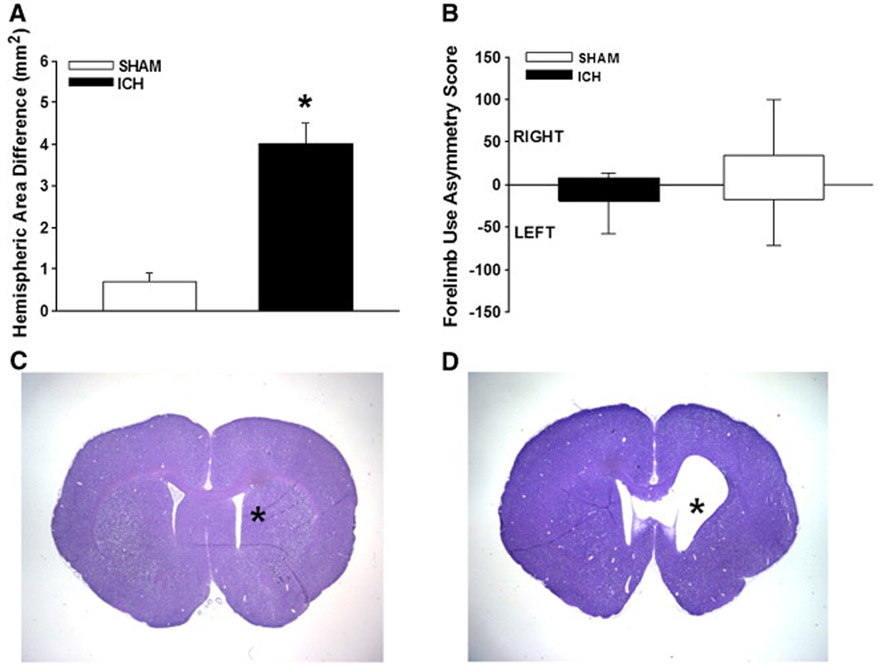
Translational neuroprotective and drug development studies need to be gauged against well-characterized functional outcomes, including motor, sensory and cognitive domains. Since intracerebral hemorrhage (ICH) causes dramatic neurological and cognitive deficits in humans, we hypothesized that ICH would result in prolonged motor-sensory and learning/memory deficits in rats. Neurological tests of sensorimotor functions were performed before ICH, 1-3 days and 10 weeks after ICH. Water maze, open field, and rotarod performance was tested 2 and 8 weeks after ICH. Early neurological evaluations revealed significant deficits, with almost full recovery by 10 weeks. The water maze revealed significant learning (but not motor) deficits at 2 weeks, but by 8 weeks, the learning deficits had diminished and significant motor deficits had emerged, coinciding with a drop in activity. The injured hemisphere showed significant atrophy at sacrifice. Therefore, ICH produced detectable cognitive and motor deficits in rats that evolved over a 10-week period, and thereby provides a suitable baseline for analysis of future therapeutic interventions following hemorrhagic stroke.
Figures
References
-
- 1999. Recommendations for standards regarding preclinical neuroprotective and restorative drug development. Stroke. 30, 2752–8. - PubMed
-
- Altumbabic M, Peeling J, Del Bigio MR, 1998. Intracerebral hemorrhage in the rat: effects of hematoma aspiration. Stroke. 29, 1917–22; discussion 1922-3. - PubMed
-
- Auriat AM, Colbourne F, 2009. Delayed rehabilitation lessens brain injury and improves recovery after intracerebral hemorrhage in rats. Brain Res. 1251, 262–8. - PubMed
-
- Bartels AL, Leenders KL, 2008. Parkinson's disease: The syndrome, the pathogenesis and pathophysiology. Cortex. - PubMed
-
- Benke T, Delazer M, Bartha L, Auer A, 2003. Basal ganglia lesions and the theory of fronto-subcortical loops: neuropsychological findings in two patients with left caudate lesions. Neurocase. 9, 70–85. - PubMed
Publication types
MeSH terms
Grants and funding
LinkOut - more resources
Full Text Sources
Medical
