. 2009 Sep;144(1-2):233-49.
doi: 10.1016/j.virusres.2009.05.006. Epub 2009 May 21.

Poxvirus protein evolution: family wide assessment of possible horizontal gene transfer events

Affiliations

Poxvirus protein evolution: family wide assessment of possible horizontal gene transfer events

Mary R Odom et al. Virus Res. 2009 Sep.

Abstract

To investigate the evolutionary origins of proteins encoded by the Poxviridae family of viruses, we examined all poxvirus protein coding genes using a method of characterizing and visualizing the similarity between these proteins and taxonomic subsets of proteins in GenBank. Our analysis divides poxvirus proteins into categories based on their relative degree of similarity to two different taxonomic subsets of proteins such as all eukaryote vs. all virus (except poxvirus) proteins. As an example, this allows us to identify, based on high similarity to only eukaryote proteins, poxvirus proteins that may have been obtained by horizontal transfer from their hosts. Although this method alone does not definitively prove horizontal gene transfer, it allows us to provide an assessment of the possibility of horizontal gene transfer for every poxvirus protein. Potential candidates can then be individually studied in more detail during subsequent investigation. Results of our analysis demonstrate that in general, proteins encoded by members of the subfamily Chordopoxvirinae exhibit greater similarity to eukaryote proteins than to proteins of other virus families. In addition, our results reiterate the important role played by host gene capture in poxvirus evolution; highlight the functions of many genes poxviruses share with their hosts; and illustrate which host-like genes are present uniquely in poxviruses and which are also present in other virus families.


Figures
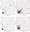
Fig. 1
Fig. 1
(A) Best BLASTP scores of poxvirus proteins against all eukaryote proteins vs. against proteins of all viruses except poxviruses. Inset shows regions described in the text. (B) Best BLASTP scores of poxvirus proteins against all bacteria proteins vs. against proteins of all viruses except poxviruses. (C) Best BLASTP scores of poxvirus proteins against all eukaryote proteins vs. against all bacteria proteins. Plotted chordopoxvirus proteins (6443 points) are represented by black squares, and plotted entomopoxvirus proteins (278 points) are represented by red squares. Notable points mentioned in the text are circled. Points circled in blue are proteins of an avian retrovirus integrated into the genome of fowlpox virus. The cluster of points circled in purple are the large subunit of ribonucleotide reductase.
Fig. 1
Fig. 1
(A) Best BLASTP scores of poxvirus proteins against all eukaryote proteins vs. against proteins of all viruses except poxviruses. Inset shows regions described in the text. (B) Best BLASTP scores of poxvirus proteins against all bacteria proteins vs. against proteins of all viruses except poxviruses. (C) Best BLASTP scores of poxvirus proteins against all eukaryote proteins vs. against all bacteria proteins. Plotted chordopoxvirus proteins (6443 points) are represented by black squares, and plotted entomopoxvirus proteins (278 points) are represented by red squares. Notable points mentioned in the text are circled. Points circled in blue are proteins of an avian retrovirus integrated into the genome of fowlpox virus. The cluster of points circled in purple are the large subunit of ribonucleotide reductase.
Fig. 2
Fig. 2
Phylogenetic reconstructions to investigate evolutionary histories of three poxvirus proteins appearing in different regions of the plot in Fig. 1A. All pictured trees were constructed by the method of Bayesian inference using MrBayes. The resulting topology for each tree agrees exactly with topology produced from the same alignment by the Maximum Likelihood method using Garli, and either agrees exactly or is very similar to topology produced by the Maximum Parsimony method using MEGA. MrBayes simulations for all three alignments were run with the GTR nucleotide substitution model and gamma distributed rate variation with an estimated proportion of invariable sites. The legend below each tree shows the scale for branch lengths as measured in expected nucleotide substitutions per site. The number to the right of each taxon name is the protein GI number for that sequence. (A) Variola virus B22R (plotted in region A near the virus axis) is a large surface glycoprotein and appears outside the poxvirus family only in the carp herpesvirus CyHV-3. (B) The interleukin-10 inhibitory cytokine (plotted on the diagonal) is evidently of eukaryote origin but has several apparent homologs in diverse virus genomes, potentially acquired in distinct gene transfer events. (C) Monoglyceride lipase (plotted in region E near the eukaryote axis) is an enzyme which may facilitate use of cellular fatty acids, and may have been acquired from a fish or reptilian host by a poxvirus ancestral to the orthopoxvirus and yatapoxvirus genera.
Fig. 3
Fig. 3
All proteins of cowpox strain GRI-90 were analyzed by taxonomic group plots, to compare the relationships of core and non-core protein subsets with proteins of eukaryotes and with proteins of viruses outside the poxvirus family. Panel (A) represents proteins classified according to genomic locus, as non-centrally located (black squares, 99 points) or centrally located (red squares, 115 points). Panel (B) represents proteins classified according to the number of poxvirus species with conserved orthologs, with genes in only 1–10 species in black (20 points), genes in 11–20 species in purple (59 points), genes in 21–30 species in blue (28 points), genes in 31–35 species in green (21 points), and genes in 36–40 species in red (86 points).
Fig. 4
Fig. 4
A genome map of cowpox strain Gri-90 is color coded (see legend) according to the degree of similarity of each cowpox protein to its best hit when compared against all virus (non-pox) or eukaryote proteins.

References

    1. Afonso C.L., Tulman E.R., Lu Z., Zsak L., Kutish G.F., Rock D.L. The genome of Fowlpox virus. J. Virol. 2000;74(8):3815–3831. - PMC - PubMed
    1. Benson D.A., Karsch-Mizrachi I., Lipman D.J., Ostell J., Wheeler D.L. GenBank. Nucleic Acids Res. 2008;36(Database issue):D25–D30. - PMC - PubMed
    1. Botstein D. A theory of modular evolution for bacteriophages. Ann. N. Y. Acad. Sci. 1980;354:484–490. - PubMed
    1. Bratke K.A., McLysaght A. Identification of multiple independent horizontal gene transfers into poxviruses using a comparative genomics approach. BMC Evol. Biol. 2008;8:67. - PMC - PubMed
    1. Condit R.C., Moussatche N., Traktman P. In a nutshell: structure and assembly of the vaccinia virion. In: Maramorosch K., Shatkin A.J., editors. vol. 66. Academic Press; 2006. pp. 31–124. (Advances in Virus Research). - PubMed

Publication types