

A comparison of brain activity evoked by single content and function words: an fMRI investigation of implicit word processing
- PMID: 19465009
- PMCID: PMC2755079
- DOI: 10.1016/j.brainres.2009.05.043
A comparison of brain activity evoked by single content and function words: an fMRI investigation of implicit word processing
Abstract
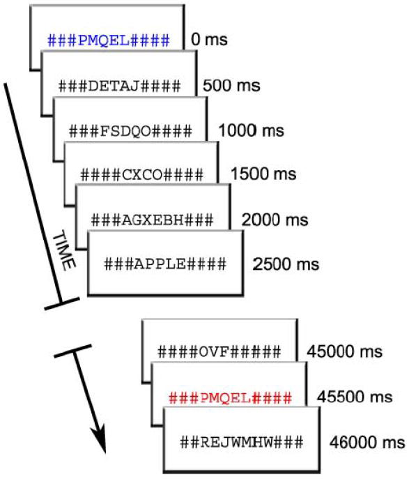
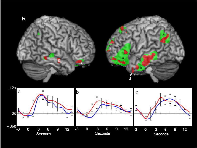
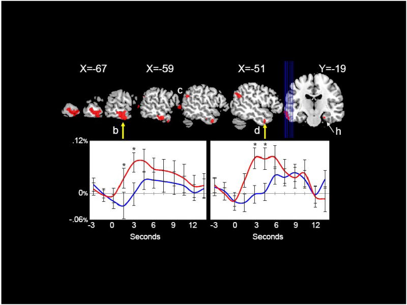
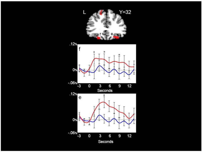
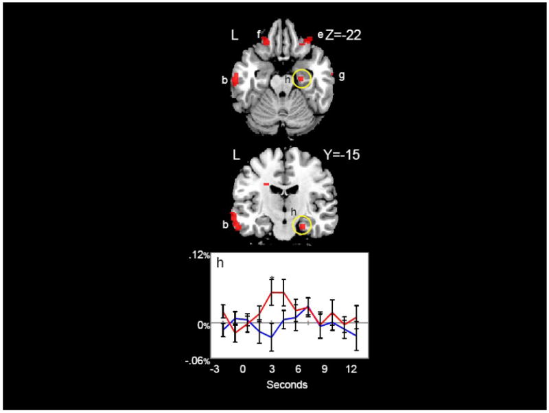
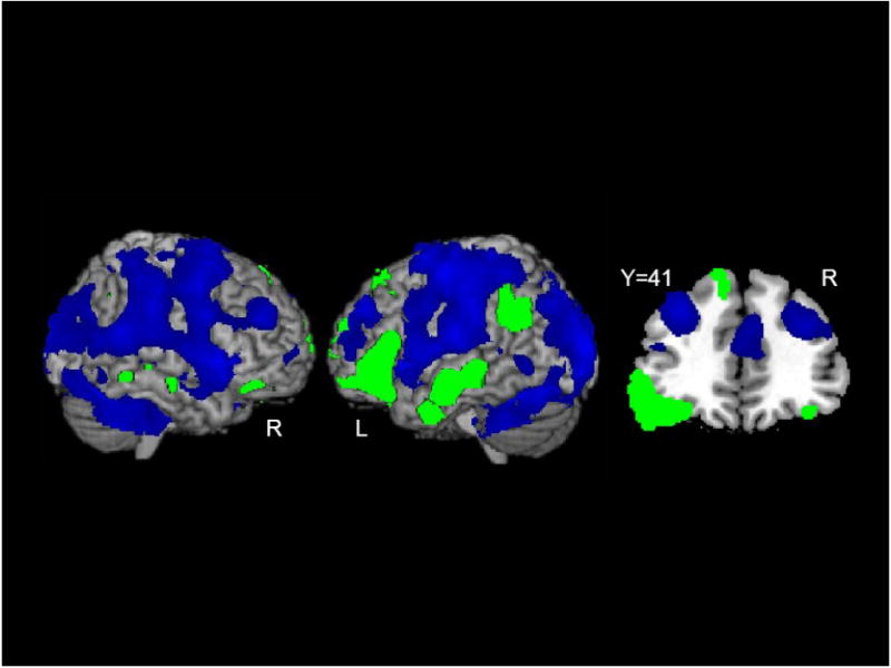
Content and function words have different roles in language and differ greatly in their semantic content. Although previous research has suggested that these different roles may be mediated by different neural substrates, the neuroimaging literature on this topic is particularly scant. Moreover, fMRI studies that have investigated differences between content and function words have utilized tasks that focus the subjects' attention on the differences between these word types. It is possible, then, that task-related differences in attention, working memory, and decision-making contribute to the differential patterns of activation observed. Here, subjects were engaged in a continuous working memory cover task while single, task-irrelevant content and function words were infrequently and irregularly presented. Nonword letter strings were displayed in black font at a fast rate (2/s). Subjects were required to either remember or retrieve occasional nonwords that were presented in colored fonts. Incidental and irrelevant to the memory task, content and function words were interspersed among nonwords at intervals of 12 to 15 s. Both word types strongly activated temporal-parietal cortex, middle and anterior temporal cortex, inferior frontal gyrus, parahippocampal gyrus, and orbital frontal cortex. Activations were more extensive in the left hemisphere. Content words elicited greater activation than function words in middle and anterior temporal cortex, a sub-region of orbital frontal cortex, and the parahippocampal region. Words also evoked extensive deactivation, most notably in brain regions previously associated with working memory and attention.
Figures
Similar articles
-
Neural representations of visual words and objects: a functional MRI study on the modularity of reading and object processing.Brain Topogr. 2007 Winter;20(2):89-96. doi: 10.1007/s10548-007-0034-1. Epub 2007 Oct 11. Brain Topogr. 2007. PMID: 17929158
-
Syllable congruency and word frequency effects on brain activation.Hum Brain Mapp. 2009 Sep;30(9):3079-88. doi: 10.1002/hbm.20730. Hum Brain Mapp. 2009. PMID: 19172625 Free PMC article.
-
Differential contributions of the anterior temporal and medial temporal lobe to the retrieval of memory for person identity information.Hum Brain Mapp. 2008 Dec;29(12):1343-54. doi: 10.1002/hbm.20469. Hum Brain Mapp. 2008. PMID: 17948885 Free PMC article.
-
Multiple systems of category learning.Neurosci Biobehav Rev. 2008;32(2):249-64. doi: 10.1016/j.neubiorev.2007.07.009. Epub 2007 Aug 15. Neurosci Biobehav Rev. 2008. PMID: 17904637 Free PMC article. Review.
-
Mechanisms underlying working memory for novel information.Trends Cogn Sci. 2006 Nov;10(11):487-93. doi: 10.1016/j.tics.2006.09.005. Epub 2006 Oct 2. Trends Cogn Sci. 2006. PMID: 17015030 Free PMC article. Review.
Cited by
-
Brief report: Anomalous neural deactivations and functional connectivity during receptive language in autism spectrum disorder: a functional MRI study.J Autism Dev Disord. 2015 Jun;45(6):1905-14. doi: 10.1007/s10803-014-2344-y. J Autism Dev Disord. 2015. PMID: 25526952 Free PMC article.
-
Grammatical Impairments in PPA.Aphasiology. 2014 Sep;28(8-9):1018-1037. doi: 10.1080/02687038.2014.912744. Aphasiology. 2014. PMID: 25642014 Free PMC article.
-
Syntax.Wiley Interdiscip Rev Cogn Sci. 2015 Mar;6(2):131-147. doi: 10.1002/wcs.1332. Epub 2014 Dec 10. Wiley Interdiscip Rev Cogn Sci. 2015. PMID: 25815105 Free PMC article. Review.
-
The role of domain-general cognitive control in language comprehension.Front Psychol. 2014 Apr 28;5:335. doi: 10.3389/fpsyg.2014.00335. eCollection 2014. Front Psychol. 2014. PMID: 24803909 Free PMC article.
-
Word meaning types acquired before vs. after age 5: implications for education.Front Psychol. 2024 Apr 5;15:1280568. doi: 10.3389/fpsyg.2024.1280568. eCollection 2024. Front Psychol. 2024. PMID: 38646119 Free PMC article.
References
-
- Andreasen NC, O’Leary DS, Cizadlo T, Arndt S, Rezai K, Watkins GL, et al. PET studies of memory: Novel versus practiced free recall of word lists. Neuroimage. 1995;2:296–305. - PubMed
-
- Andrews S. The effect of orthographic similarity on lexical retrieval: Resolving neighborhood conflicts. Psychonomic Bulletin and Review. 1997;4:439–461.
-
- Bechara A, Damasio H, Damasio AR. Emotion, decision making and the orbitofrontal cortex. Cerebral Cortex. 2000;10(3):295–307. - PubMed
-
- Beckman CF, Jenkinson M, Smith SM. General multi-level linear modelling for group analysis in FMRI. Neuroimage. 2003;20:1052–1063. - PubMed
Publication types
MeSH terms
Grants and funding
LinkOut - more resources
Full Text Sources
Medical
