. 2009 Jul 24;284(30):20329-39.
doi: 10.1074/jbc.M109.010264. Epub 2009 May 22.

TDP-43 is intrinsically aggregation-prone, and amyotrophic lateral sclerosis-linked mutations accelerate aggregation and increase toxicity

Affiliations

TDP-43 is intrinsically aggregation-prone, and amyotrophic lateral sclerosis-linked mutations accelerate aggregation and increase toxicity

Brian S Johnson et al. J Biol Chem. .

Erratum in

  • J Biol Chem. 2009 Sep 11;284(37):25459

Abstract

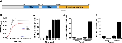
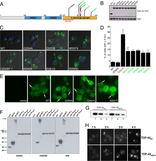
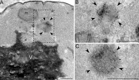
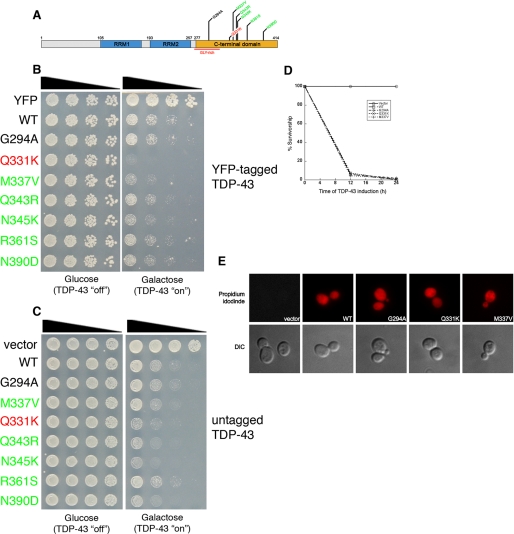
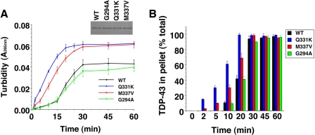
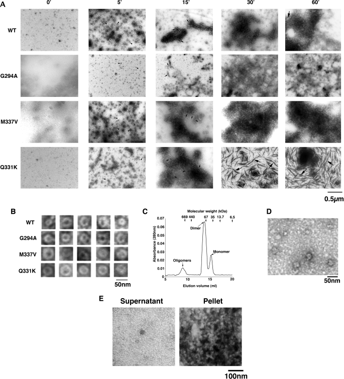
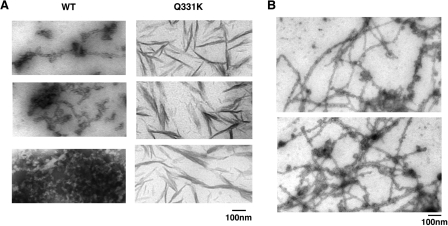
Non-amyloid, ubiquitinated cytoplasmic inclusions containing TDP-43 and its C-terminal fragments are pathological hallmarks of amyotrophic lateral sclerosis (ALS), a fatal motor neuron disorder, and frontotemporal lobar degeneration with ubiquitin-positive inclusions (FTLD-U). Importantly, TDP-43 mutations are linked to sporadic and non-SOD1 familial ALS. However, TDP-43 is not the only protein in disease-associated inclusions, and whether TDP-43 misfolds or is merely sequestered by other aggregated components is unclear. Here, we report that, in the absence of other components, TDP-43 spontaneously forms aggregates bearing remarkable ultrastructural similarities to TDP-43 deposits in degenerating neurons of ALS and FTLD-U patients [corrected] . The C-terminal domain of TDP-43 is critical for spontaneous aggregation. Several ALS-linked TDP-43 mutations within this domain (Q331K, M337V, Q343R, N345K, R361S, and N390D) increase the number of TDP-43 aggregates and promote toxicity in vivo. Importantly, mutations that promote toxicity in vivo accelerate aggregation of pure TDP-43 in vitro. Thus, TDP-43 is intrinsically aggregation-prone, and its propensity for toxic misfolding trajectories is accentuated by specific ALS-linked mutations.


Figures

FIGURE 1.
FIGURE 1.
Aggregation of TDP-43 and TDP-43 fragments in vitro. A, a diagram of the domain structure of TDP-43 indicating both RNA recognition motifs (RRM1 and RRM2) and the glycine-rich C-terminal domain. B, TDP-43 or the indicated TDP-43 fragment (1–275 or 188–414) (3 μm) were incubated at 25 °C with agitation for 0–120 min. The extent of aggregation was determined by turbidity. Values represent means ± S.D. (n = 3). C, aggregation of WT TDP-43 as in B assessed by sedimentation analysis. Values represent means ± S.D. (n = 3). D, His-TDP-43 or Sup35-NM (5 μm) was incubated for either 0 min (soluble protein) or 2 h (aggregated protein) at 25 °C with agitation. Reactions were then processed for Congo Red Binding. Values represent means ± S.D. (n = 3). E, TDP-43 or Sup35-NM (5 μm) was incubated for either 0 min (soluble protein) or 2 h (aggregated protein) at 25 °C with agitation. Reactions were then processed for Thioflavin-T fluorescence. Values represent means ± S.D. (n = 3).
FIGURE 2.
FIGURE 2.
Aggregation of TDP-43 and ALS-linked mutants in vivo. A, schematic indicating disease-associated TDP-43 mutations. Color code of mutations indicates aggregation compared with WT (red = considerably more aggregates than WT, green = more aggregates than WT, and black = as many aggregates as WT). B, YFP alone, WT, and mutant TDP-43-YFP expression levels were determined by immunoblotting with an anti-GFP antibody. Phosphoglycerate kinase 1 (Pgk1) was used as a loading control. C, representative fluorescent microscopy images of 4′,6-diamidino-2-phenylindole-stained (blue to denote the position of the nucleus) WT or mutant TDP-43-YFP (green). D, the effect of TDP-43 mutations on aggregation in vivo was quantified by counting the number of cells containing >3 foci. Values represent means ± S.E. (n ≥ 3, at least 200 cells per sample). Cells expressing YFP alone did not contain foci (data not shown). E, untagged TDP-43 also forms aggregates in yeast cells. Immunocytochemistry with an anti-TDP-43 antibody was used to visualize TDP-43 expression. Arrows point to cytoplasmic TDP-43 inclusions. Cells transformed with an empty vector were used as a control for antibody specificity. F, sedimentation assay to demonstrate TDP-43 forms insoluble aggregates in yeast cells. Yeast cells expressing YFP, htt25QCFP, htt103Q-CFP, or WT and mutant TDP-43-YFP for 6 h were lysed and processed for sedimentation assay as in a previous study (19). Soluble and insoluble fractions were separated by centrifugation. 20% of the pellet fraction and 10% of soluble and total fractions were resolved by SDS-PAGE followed by immunoblotting with anti-GFP antibody. Whereas YFP was completely soluble, WT and TDP-43 partitioned into soluble and insoluble fractions, similar to the aggregation-prone fragments of the huntingtin protein (htt25Q (mostly soluble) or htt103Q (mostly insoluble)). The arrow indicates the top of gel. G, sedimentation assay performed as in F except that WT and Q331K TDP-43 constructs were induced for 4 h. At this time point, a greater relative amount of Q331K is present in the insoluble pellet fraction than WT TDP-43. T, total; S, soluble; P, pellet. Representative image from three separate experiments is shown. H, time-course analysis of WT and Q331K TDP-43 aggregation demonstrates Q331K forms more discrete aggregates than WT at early time points.
FIGURE 3.
FIGURE 3.
EM of TDP-43 aggregates in yeast. A–C, low magnification TEM shows an example of a granular aggregate in yeast cells expressing the Q331K TDP-43 mutant (boxed region (A), high magnification +25° tilt view of boxed region in A (B), and additional high magnification view of TDP-43 aggregate (C)). m = mitochondria; V = vacuole; arrowheads delineate TDP-43 aggregate; bars = 0.25 μm.
FIGURE 4.
FIGURE 4.
The effect of ALS-linked TDP-43 mutations on toxicity. A, schematic indicating disease-associated TDP-43 mutations shown above. Color code of mutations indicates toxicity compared with WT (red = considerably more toxic than WT; green = slightly more toxic than WT; black = as toxic as WT). B, spotting assay to compare the toxicity of WT and mutant TDP-43. Serial dilutions of yeast cells transformed with galactose-inducible YFP, WT, or mutant TDP-43-YFP constructs. Transformants were spotted on glucose- (non-inducing) or galactose- (inducing) containing agar plates, and growth assessed after 48–72 h. C, spotting assay to compare the toxicity of WT and mutant TDP-43. Serial dilutions of yeast cells transformed with galactose-inducible untagged WT or mutant TDP-43 constructs. Transformants were spotted on glucose- (non-inducing) or galactose- (inducing) containing agar plates, and growth was assessed after 48–72 h. D, survivorship curve during TDP-43 induction, using the high copy 2μ vector. After induction of empty vector, TDP-43 WT, G294A, Q331K, or M337V, survivorship was determined at the indicated time points by harvesting cells at A600 nm = 1, diluting 1:1000, and plating 300 μl of these cells onto synthetic media containing 2% glucose (represses TDP-43 expression). Plates were incubated at 30 °C, and colony forming units were determined after 2 days. E, TDP-43 expression causes cell death. TDP-43 expression was induced for 6 h, and cells were stained with propidium iodide (PI) to assess viability. WT and mutant TDP-43-expressing cells were positive for PI staining (indicating cell death), whereas empty vector-containing cells were negative for PI staining (indicating viability).
FIGURE 5.
FIGURE 5.
Q331K and M337V accelerate spontaneous TDP-43 aggregation. A and B, TDP-43 or the indicated ALS-linked TDP-43 mutant (Q331K, M337V, or G294A) (3 μm) were incubated at 25 °C with agitation for 0–60 min. The extent of aggregation was determined by turbidity (A) or sedimentation analysis (B). Values represent means ± S.D. (n = 3). Inset (A) the equal input of TDP-43 proteins is shown on a Coomassie-stained gel.
FIGURE 6.
FIGURE 6.
Pure TDP-43 and ALS-linked mutant aggregates resemble TDP-43 aggregates in degenerating neurons of ALS patients. A, TDP-43 or the indicated ALS-linked TDP-43 mutant (Q331K, M337V, or G294A) (3 μm) were incubated at 25 °C with agitation for 0–60 min. At various times, reactions were processed for EM. Arrowheads denote small oligomers, small arrows denote filament-like structures, and large arrows denote thread-like structures. Bar, 0.5 μm. B, gallery of TDP-43 and ALS-linked TDP-43 mutant oligomers formed after 5 min. Bar, 50 nm. C, TDP-43 was incubated at 25 °C with agitation for 5 min and processed for Superdex 200 size-exclusion chromatography. TDP-43 eluted as a predominantly dimeric and monomeric species, but a small fraction eluted as oligomeric species. Arrows at the top indicate elution volumes of the molecular weight standards. D, EM of oligomeric pool eluted from the Superdex 200 column. Bar, 50 nm. E, TDP-43 (3 μm) was incubated at 25 °C with agitation for 30 min. Supernatant and pellet fractions were separated and processed for EM. Bar, 100 nm.
FIGURE 7.
FIGURE 7.
Thread-like aggregates formed by TDP-43. A, gallery of thread-like aggregates of TDP-43 and Q331K formed after 60 min of agitation at 25 °C. Bar, 100 nm. B, longer and more regular granulo-filamentous forms populated by WT TDP-43 after 2 h of agitation at 25 °C. Bar, 100 nm.

References

    1. Ayala Y. M., Pantano S., D'Ambrogio A., Buratti E., Brindisi A., Marchetti C., Romano M., Baralle F. E. (2005) J. Mol. Biol. 348, 575–588 - PubMed
    1. Ayala Y. M., Misteli T., Baralle F. E. (2008) Proc. Natl. Acad. Sci. U.S.A. 105, 3785–3789 - PMC - PubMed
    1. Buratti E., Baralle F. E. (2001) J. Biol. Chem. 276, 36337–36343 - PubMed
    1. Neumann M., Sampathu D. M., Kwong L. K., Truax A. C., Micsenyi M. C., Chou T. T., Bruce J., Schuck T., Grossman M., Clark C. M., McCluskey L. F., Miller B. L., Masliah E., Mackenzie I. R., Feldman H., Feiden W., Kretzschmar H. A., Trojanowski J. Q., Lee V. M. (2006) Science 314, 130–133 - PubMed
    1. Forman M. S., Trojanowski J. Q., Lee V. M. (2007) Curr. Opin. Neurobiol. 17, 548–555 - PMC - PubMed

Publication types

MeSH terms