. 2009 Jul 17;284(29):19420-6.
doi: 10.1074/jbc.M901744200. Epub 2009 May 22.

Interleukin-33 is biologically active independently of caspase-1 cleavage

Affiliations

Interleukin-33 is biologically active independently of caspase-1 cleavage

Dominique Talabot-Ayer et al. J Biol Chem. .

Abstract

The new interleukin (IL)-1 family cytokine IL-33 is synthesized as a 30-kDa precursor. Like pro-IL-1beta, human pro-IL-33 was reported to be cleaved by caspase-1 to generate an 18-kDa fragment, which is sufficient to activate signaling by the IL-33 receptor T1/ST2. However, the proposed caspase-1 cleavage site is poorly conserved between species. In addition, it is not clear whether caspase-1 cleavage of pro-IL-33 occurs in vivo and whether, as for IL-1beta, this cleavage is a prerequisite for IL-33 secretion and bioactivity. In this study, we further investigated caspase-1 cleavage of mouse and human pro-IL-33 and assessed the potential bioactivity of the IL-33 precursor. We observed the generation of a 20-kDa IL-33 fragment in cell lysates, which was enhanced by incubation with caspase-1. However, in vitro assays of mouse and human pro-IL-33 indicated that IL-33 is not a direct substrate for this enzyme. Consistently, caspase-1 activation in THP-1 cells induced cleavage of pro-IL-1beta but not of pro-IL-33, and activated THP-1 cells released full-length pro-IL-33 into culture supernatants. Finally, addition of full-length pro-IL-33 induced T1/ST2-dependent IL-6 secretion in mast cells. However, we observed in situ processing of pro-IL-33 in mast cell cultures, and it remains to be determined whether full-length pro-IL-33 itself indeed represents the bioactive species. In conclusion, our data indicate that pro-IL-33 is not a direct substrate for caspase-1. In addition, our results clearly show that caspase-1 cleavage is not required for pro-IL-33 secretion and bioactivity, highlighting major differences between IL-1beta and IL-33.


Figures

FIGURE 1.
FIGURE 1.
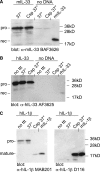
Generation of a 20-kDa fragment of IL-33 in cell lysates is enhanced in the presence of caspase-1. A, total cell lysates of 293T cells overexpressing mouse pro-IL-33 (mIL-33) or of control 293T cells transfected with empty vector (control) were either left untreated (no ttt) or incubated for 6 h at 37 °C in the presence (Csp 37°) or absence (37°) of recombinant human active caspase-1. Samples were analyzed by Western blotting with a polyclonal anti-mIL-33 antibody (BAF3626). B, total cell lysates of 293T cells overexpressing D88A mutant mouse pro-IL-33, D106A mutant mouse pro-IL-33, or wild-type mouse pro-IL-33 (WT) were incubated for 6 h at 37 °C in the absence or presence of recombinant human active caspase-1. C, total cell lysates of 293T cells overexpressing human pro-IL-33 (hIL-33) or of control 293T cells transfected with empty vector (control) were either left untreated or incubated for 6 h at 37 °C in the absence or presence of recombinant human active caspase-1. Samples were analyzed by Western blotting with a polyclonal anti-hIL-33 antibody (AF3625). A–C, the positions of full-length pro-IL-33 (pro), rIL-33 (rec), and the 20-kDa fragment (arrow) are indicated on the left. Molecular mass markers are indicated on the right. D, total cell lysates of 293T cells overexpressing human pro-IL-1β (hIL-1β) were incubated for 6 h at 37 °C in the absence or presence of recombinant human active caspase-1. Samples were analyzed by Western blotting with the D116 antibody, which specifically recognizes the cleaved 17-kDa form of IL-1β (arrow).
FIGURE 2.
FIGURE 2.
IL-33 is not a direct substrate for caspase-1. A, in vitro synthesized mouse pro-IL-33 (mIL-33) or a control in vitro synthesis reaction performed without cDNA (no DNA) was incubated for 6 h at 37 °C in the absence (37°) or presence (Csp 37°) of recombinant human active caspase-1. Samples were analyzed by Western blotting with a polyclonal anti-mIL-33 antibody (BAF3626). B, in vitro synthesized human pro-IL-33 (hIL-33) or a control in vitro synthesis reaction performed without cDNA was either left untreated (no ttt) or incubated for 6 h at 37 °C in the absence or presence of recombinant human active caspase-1. Samples were analyzed by Western blotting with a polyclonal anti-hIL-33 antibody (AF3625). A and B, the positions of full-length pro-IL-33 (pro) and rIL-33 (rec) are indicated on the left. Molecular size markers are indicated on the right. C, in vitro synthesized human pro-IL-1β (hIL-1β) was either left untreated or incubated for 6 h at 37 °C in the absence or presence of recombinant human active caspase-1. Samples were analyzed by Western blotting with the MAB201 antibody, which recognizes both pro-IL-1β and cleaved IL-1β (left panel) and with the D116 antibody, which specifically recognizes the cleaved 17-kDa form of IL-1β (right panel).
FIGURE 3.
FIGURE 3.
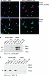
IL-33 is released in its precursor form by LPS-activated THP-1 cells. A, localization of IL-33 in PMA- and LPS-stimulated THP-1 cells expressing GFP alone (THP-1 pWPI, left panels) or human pro-IL-33 and GFP (THP-1 pWPI-hIL-33, right panels) was analyzed by immunocytochemistry (upper panels). Anti-IL-33 staining (red) was essentially detected in cell nuclei, which were visualized with 4′,6-diamidino-2-phenylindole (DAPI; blue). Efficiency of lentiviral transduction was verified by monitoring GFP expression (lower panels). B, THP-1 cells expressing GFP (pWPI) or human pro-IL-33 and GFP (IL-33) were primed with PMA and stimulated (LPS) or not (Ø) with LPS for 6 h. Secreted (supernatant) and intracellular (lysate) proteins were analyzed by Western blotting using a polyclonal anti-hIL-33 antibody (AF3625). The positions of full-length pro-IL-33 (pro), rIL-33 (rec), and the 20-kDa fragment (arrow) are indicated on the left. Molecular mass markers are indicated on the right. C, the same samples as in B were analyzed by Western blotting with the D116 antibody, which specifically recognizes the cleaved 17-kDa form of IL-1β (arrow).
FIGURE 4.
FIGURE 4.
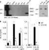
Purified pro-IL-33 exerts T1/ST2-dependent biological effects on mast cells. A, mouse pro-IL-33 was purified from total cell lysates or nuclear extracts of transfected 293T cells using sST2-Fc/protein G beads, as described under “Experimental Procedures.” Total cell lysate used for purification (load, 10 μl of 500 μl), column flow-through (FT, 10 μl of 500 μl), first wash (wash, 10 μl of 500), and eluted fractions 1–4 (el1–el4, 10 μl of 100 μl) were analyzed by Western blotting using the Nessy-1 anti-IL-33 antibody (left panel). Alternatively, 10 μl of ST2-Fc/protein G beads was incubated with 20 μl of total cell lysate and washed, and pro-IL-33 was eluted directly in Laemmli buffer (lb el, right panel). lysate indicates the total cell lysate (1.5 μl). B, wild-type (WT) and T1/ST2 knock-out (ST2KO) BMMC were stimulated for 24 h with 10 μl of purified pro-IL-33, 1 μg/ml LPS, or 1 ng/ml rmIL-33 before IL-6 secretion was assessed in culture supernatants by enzyme-linked immunosorbent assay. Results shown are mean ± S.E. of culture triplicates. Similar results were obtained with eluates from three independent purifications. *, p < 0.05 versus control; &, p < 0.05 versus wild-type BMMC with the same stimulation.
FIGURE 5.
FIGURE 5.
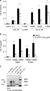
Bioactivity of in vitro synthesized full-length pro-IL-33. A, P815 mastocytoma cells were stimulated for 24 h with indicated doses of rmIL-33, with the indicated dilutions of in vitro synthesized mouse pro-IL-33, or with a control in vitro synthesis reaction performed without cDNA (no DNA) in the presence or absence of sST2-Fc (50 μg/ml) before IL-6 secretion was assessed in culture supernatants by enzyme-linked immunosorbent assay. B, P815 mastocytoma cells were stimulated for 24 h with the indicated dilutions of in vitro synthesized mouse pro-IL-33 mutant D88A or D106A in the presence or absence of sST2-Fc (50 μg/ml) before IL-6 secretion was assessed in culture supernatants by enzyme-linked immunosorbent assay. A and B, results shown are the mean ± S.E. of triplicate cultures in a representative experiment. *, p < 0.05 versus control; &, p < 0.05 versus the same stimulation in the absence of sST2-Fc. C, P815 mastocytoma cells were incubated for 24 h in Opti-MEM without or with 5 μl (↓ pro-IL-33 5) or 2 μl (↓ pro-IL-33 2) of in vitro synthesized mouse pro-IL-33 or with 5 μl of a control in vitro synthesis reaction performed without cDNA (↓ no DNA). Culture supernatants were harvested, methanol-precipitated, resuspended in Laemmli buffer, and analyzed by Western blotting with a polyclonal anti-mIL-33 antibody (BAF3626). In this experiment, full-length pro-IL-33 was almost completely processed to yield fragments of lower molecular mass. pro-IL-33 indicates total cell lysate of mouse pro-IL-33-expressing 293T cells. The positions of full-length pro-IL-33 (pro), rIL-33 (rec), and 28- and 20-kDa fragments (arrows) are indicated on the left. Molecular mass markers are indicated on the right.

References

    1. Schmitz J., Owyang A., Oldham E., Song Y., Murphy E., McClanahan T. K., Zurawski G., Moshrefi M., Qin J., Li X., Gorman D. M., Bazan J. F., Kastelein R. A. (2005) Immunity 23, 479–490 - PubMed
    1. Howard A. D., Kostura M. J., Thornberry N., Ding G. J., Limjuco G., Weidner J., Salley J. P., Hogquist K. A., Chaplin D. D., Mumford R. A., Schmidt J. A., Tocci M. J. (1991) J. Immunol. 147, 2964–2969 - PubMed
    1. Lüschen S., Ussat S., Krönke M., Adam-Klages S. (1998) Biochem. Biophys. Res. Commun. 253, 92–98 - PubMed
    1. Lamkanfi M., Kanneganti T. D., Van Damme P., Vanden Berghe T., Vanoverberghe I., Vandekerckhove J., Vandenabeele P., Gevaert K., Núñez G. (2008) Mol. Cell. Proteomics 7, 2350–2363 - PMC - PubMed
    1. Carriere V., Roussel L., Ortega N., Lacorre D. A., Americh L., Aguilar L., Bouche G., Girard J. P. (2007) Proc. Natl. Acad. Sci. U.S.A. 104, 282–287 - PMC - PubMed

Publication types

MeSH terms