

Sustained hypoxia promotes the development of a pulmonary artery-specific chronic inflammatory microenvironment
- PMID: 19465514
- PMCID: PMC2742800
- DOI: 10.1152/ajplung.90591.2008
Sustained hypoxia promotes the development of a pulmonary artery-specific chronic inflammatory microenvironment
Abstract
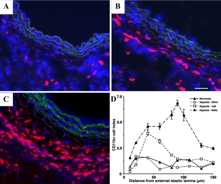
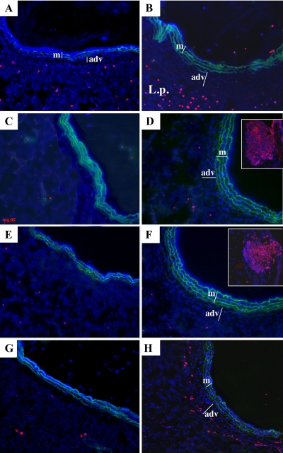
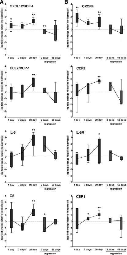
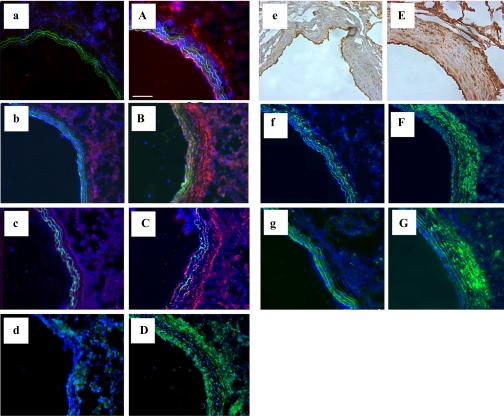
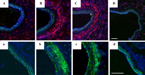
Recent studies demonstrate that sustained hypoxia induces the robust accumulation of leukocytes and mesenchymal progenitor cells in pulmonary arteries (PAs). Since the factors orchestrating hypoxia-induced vascular inflammation are not well-defined, the goal of this study was to identify mediators potentially responsible for recruitment to and retention and differentiation of circulating cells within the hypoxic PA. We analyzed mRNA expression of 44 different chemokine/chemokine receptor, cytokine, adhesion, and growth and differentiation genes in PAs obtained via laser capture microdissection in adjacent lung parenchyma and in systemic arteries by RT-PCR at several time points of hypoxic exposure (1, 7, and 28 days) in Wistar-Kyoto rats. Analysis of inflammatory cell accumulation and protein expression of selected genes was concomitantly assessed by immunochemistry. We found that hypoxia induced progressive accumulation of monocytes and dendritic cells in the vessel wall with few T cells and no B cells or neutrophils. Upregulation of stromal cell-derived factor-1 (SDF-1), VEGF, growth-related oncogene protein-alpha (GRO-alpha), C5, ICAM-1, osteopontin (OPN), and transforming growth factor-beta (TGF-beta) preceded mononuclear cell influx. With time, a more complex pattern of gene expression developed with persistent upregulation of adhesion molecules (ICAM-1, VCAM-1, and OPN) and monocyte/fibrocyte growth and differentiation factors (TGF-beta, endothelin-1, and 5-lipoxygenase). On return to normoxia, expression of many genes (including SDF-1, monocyte chemoattractant protein-1, C5, ICAM-1, and TGF-beta) rapidly returned to control levels, changes that preceded the disappearance of monocytes and reversal of vascular remodeling. In conclusion, sustained hypoxia leads to the development of a complex, PA-specific, proinflammatory microenvironment capable of promoting recruitment, retention, and differentiation of circulating monocytic cell populations that contribute to vascular remodeling.
Figures

Similar articles
-
Hypoxia, leukocytes, and the pulmonary circulation.J Appl Physiol (1985). 2005 Feb;98(2):715-21. doi: 10.1152/japplphysiol.00840.2004. J Appl Physiol (1985). 2005. PMID: 15649883 Review.
-
Emergence of fibroblasts with a proinflammatory epigenetically altered phenotype in severe hypoxic pulmonary hypertension.J Immunol. 2011 Sep 1;187(5):2711-22. doi: 10.4049/jimmunol.1100479. Epub 2011 Aug 3. J Immunol. 2011. PMID: 21813768 Free PMC article.
-
Monocytes and dendritic cells in a hypoxic environment: Spotlights on chemotaxis and migration.Immunobiology. 2008;213(9-10):733-49. doi: 10.1016/j.imbio.2008.07.031. Epub 2008 Sep 21. Immunobiology. 2008. PMID: 18926289 Review.
-
Impact of interleukin-6 on hypoxia-induced pulmonary hypertension and lung inflammation in mice.Respir Res. 2009 Jan 27;10(1):6. doi: 10.1186/1465-9921-10-6. Respir Res. 2009. PMID: 19173740 Free PMC article.
-
Hypoxia-induced pulmonary vascular remodeling requires recruitment of circulating mesenchymal precursors of a monocyte/macrophage lineage.Am J Pathol. 2006 Feb;168(2):659-69. doi: 10.2353/ajpath.2006.050599. Am J Pathol. 2006. PMID: 16436679 Free PMC article.
Cited by
-
Hypoxia and aging: molecular mechanisms, diseases, and therapeutic targets.MedComm (2020). 2024 Oct 15;5(11):e786. doi: 10.1002/mco2.786. eCollection 2024 Nov. MedComm (2020). 2024. PMID: 39415849 Free PMC article. Review.
-
Alveolar hypoxia promotes murine lung tumor growth through a VEGFR-2/EGFR-dependent mechanism.Cancer Prev Res (Phila). 2012 Aug;5(8):1061-71. doi: 10.1158/1940-6207.CAPR-12-0069-T. Epub 2012 Jun 14. Cancer Prev Res (Phila). 2012. PMID: 22700853 Free PMC article.
-
Targeting RUNX1 as a novel treatment modality for pulmonary arterial hypertension.Cardiovasc Res. 2022 Dec 29;118(16):3211-3224. doi: 10.1093/cvr/cvac001. Cardiovasc Res. 2022. PMID: 35018410 Free PMC article.
-
Dynamic and diverse changes in the functional properties of vascular smooth muscle cells in pulmonary hypertension.Cardiovasc Res. 2018 Mar 15;114(4):551-564. doi: 10.1093/cvr/cvy004. Cardiovasc Res. 2018. PMID: 29385432 Free PMC article. Review.
-
Plexiform Arteriopathy in Rodent Models of Pulmonary Arterial Hypertension.Am J Pathol. 2019 Jun;189(6):1133-1144. doi: 10.1016/j.ajpath.2019.02.005. Epub 2019 Mar 26. Am J Pathol. 2019. PMID: 30926336 Free PMC article. Review.
References
-
- Betsuyaku T, Griffin GL, Watson MA, Senior RM. Laser capture microdissection and real-time reverse transcriptase/polymerase chain reaction of bronchiolar epithelium after bleomycin. Am J Respir Cell Mol Biol 25: 278–284, 2001. - PubMed
-
- Ceradini DJ, Kulkarni AR, Callaghan MJ, Tepper OM, Bastidas N, Kleinman ME, Capla JM, Galiano RD, Levine JP, Gurtner GC. Progenitor cell trafficking is regulated by hypoxic gradients through HIF-1 induction of SDF-1. Nat Med 10: 858–864, 2004. - PubMed
-
- Chen YF, Feng JA, Li P, Xing D, Zhang Y, Serra R, Ambalavanan N, Majid-Hassan E, Oparil S. Dominant negative mutation of the TGF-β receptor blocks hypoxia-induced pulmonary vascular remodeling. J Appl Physiol 100: 564–571, 2006. - PubMed
-
- Chin KM, Rubin LJ. Pulmonary arterial hypertension. J Am Coll Cardiol 51: 1527–1538, 2008. - PubMed
Publication types
MeSH terms
Substances
Grants and funding
LinkOut - more resources
Full Text Sources
Medical
Molecular Biology Databases
Research Materials
Miscellaneous