. 2009 Aug;297(2):L347-61.
doi: 10.1152/ajplung.90559.2008. Epub 2009 May 22.

The contribution of inositol 1,4,5-trisphosphate and ryanodine receptors to agonist-induced Ca(2+) signaling of airway smooth muscle cells

Affiliations

The contribution of inositol 1,4,5-trisphosphate and ryanodine receptors to agonist-induced Ca(2+) signaling of airway smooth muscle cells

Yan Bai et al. Am J Physiol Lung Cell Mol Physiol. 2009 Aug.

Abstract

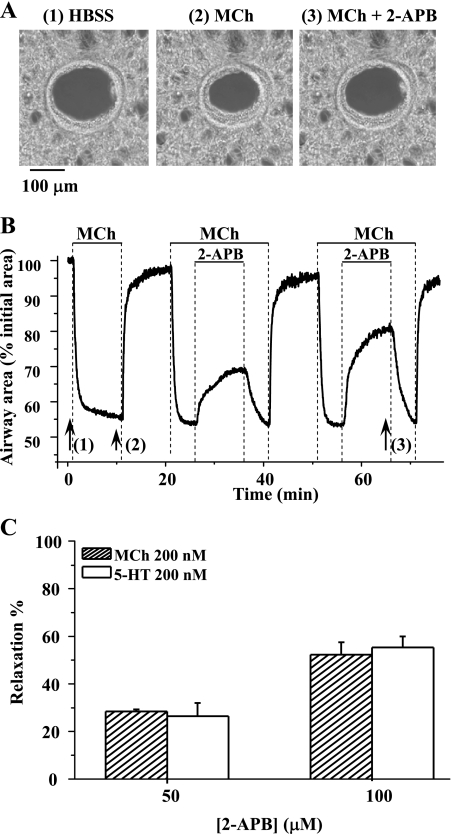
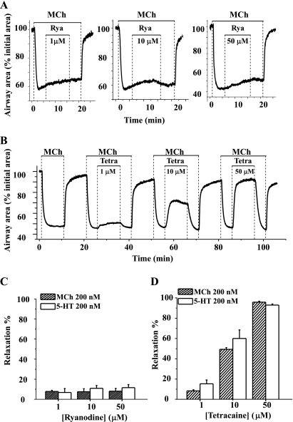
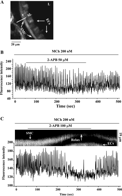
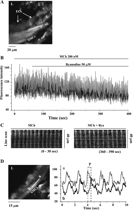
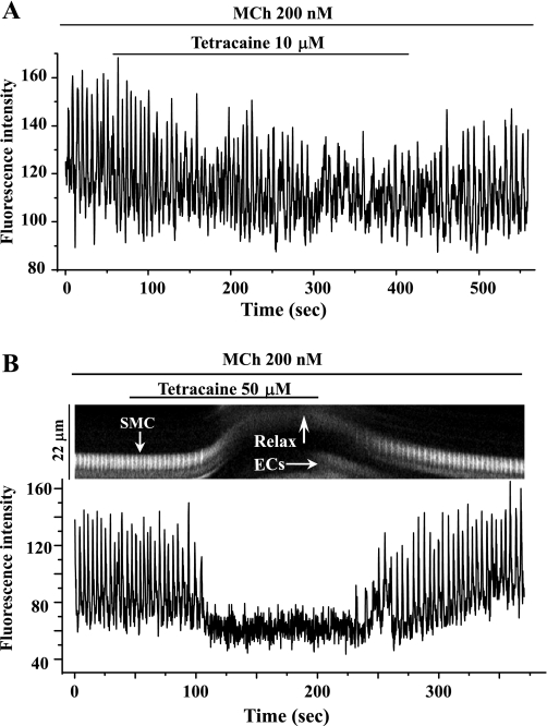
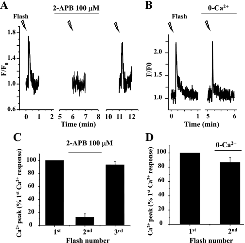
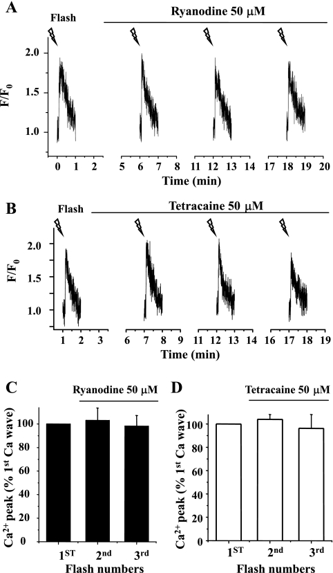
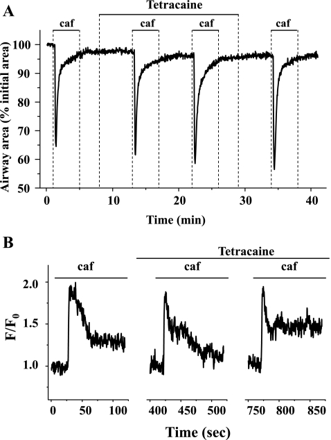
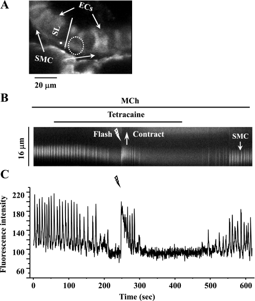
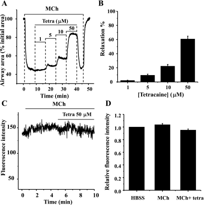
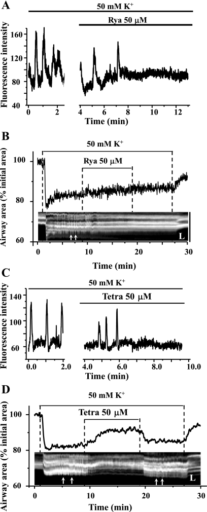
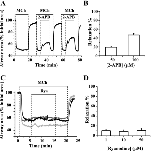
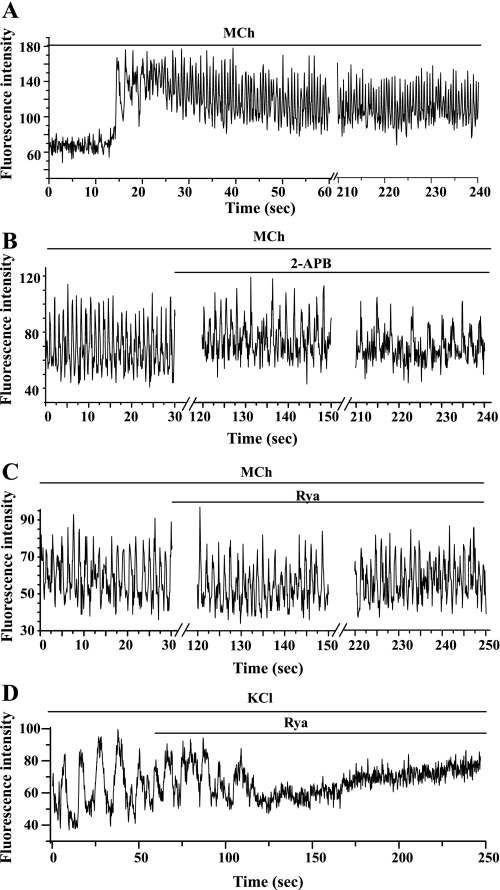
The relative contribution of inositol 1,4,5-trisphosphate (IP(3)) receptors (IP(3)Rs) and ryanodine receptors (RyRs) to agonist-induced Ca(2+) signaling in mouse airway smooth muscle cells (SMCs) was investigated in lung slices with phase-contrast or laser scanning microscopy. At room temperature (RT), methacholine (MCh) or 5-hydroxytryptamine (5-HT) induced Ca(2+) oscillations and an associated contraction in small airway SMCs. The subsequent exposure to an IP(3)R antagonist, 2-aminoethoxydiphenyl borate (2-APB), inhibited the Ca(2+) oscillations and induced airway relaxation in a concentration-dependent manner. 2-APB also inhibited Ca(2+) waves generated by the photolytic release of IP(3). However, the RyR antagonist ryanodine had no significant effect, at any concentration, on airway contraction or agonist- or IP(3)-induced Ca(2+) oscillations or Ca(2+) wave propagation. By contrast, a second RyR antagonist, tetracaine, relaxed agonist-contracted airways and inhibited agonist-induced Ca(2+) oscillations in a concentration-dependent manner. However, tetracaine did not affect IP(3)-induced Ca(2+) release or wave propagation nor the Ca(2+) content of SMC Ca(2+) stores as evaluated by Ca(2+)-release induced by caffeine. Conversely, both ryanodine and tetracaine completely blocked agonist-independent slow Ca(2+) oscillations induced by KCl. The inhibitory effects of 2-APB and absence of an effect of ryanodine on MCh-induced airway contraction or Ca(2+) oscillations of SMCs were also observed at 37 degrees C. In Ca(2+)-permeable SMCs, tetracaine inhibited agonist-induced contraction without affecting intracellular Ca(2+) levels indicating that relaxation also resulted from a reduction in Ca(2+) sensitivity. These results indicate that agonist-induced Ca(2+) oscillations in mouse small airway SMCs are primary mediated via IP(3)Rs and that tetracaine induces relaxation by both decreasing Ca(2+) sensitivity and inhibiting agonist-induced Ca(2+) oscillations via an IP(3)-dependent mechanism.


Figures

Fig. 1.
Fig. 1.
The effect of 2-aminoethoxydiphenyl borate (2-APB) on agonist-induced airway contraction. A: phase-contrast images of the same airway (at times indicated by numbered arrows in B) showing 1) the relaxed initial state of the airway, 2) the contractile state of the airway induced by 200 nM methacholine (MCh), and 3) the relaxation induced by 100 μM 2-APB. B: the change in airway area with respect to time showing the airway contraction in response to 200 nM MCh and the subsequent relaxation induced by either 50 or 100 μM 2-APB. The magnitude of airway relaxation increased as the concentration of 2-APB increased. C: summary of the relaxation induced in airways contracted with 200 nM MCh or 200 nM 5-hydroxytryptamine (5-HT) by 50 and 100 μM 2-APB. The magnitude of the relaxation was measured after 10 min of 2-APB exposure and is expressed as a percentage of the contracted luminal area induced by agonists. Each column represents the mean ± SE from 6 different experiments on different slices from 3 mice.
Fig. 2.
Fig. 2.
Effects of ryanodine and tetracaine on agonist-induced airway contraction. Representative experiments showing airway contraction in response to 200 nM MCh and the subsequent responses to different concentrations of ryanodine (Rya; A; 1, 10, or 50 μM) or tetracaine (Tetra; B; 1, 10, or 50 μM) are shown. At all concentrations tested, ryanodine had no obvious effect on MCh-induced contraction. Because the effects of ryanodine are irreversible, 3 individual lung slices were used to test the effects of each concentration of ryanodine. By contrast, tetracaine relaxed MCh-contracted airways in a concentration-dependent manner. Because the effects of tetracaine are reversible, the same lung slice was used to examine the effects of multiple concentrations of tetracaine. A summary of the airway responses to ryanodine (C) and tetracaine (D) in the presence of MCh or 5-HT is shown. Relaxation was measured after 10 min of exposure to ryanodine or tetracaine and is expressed as a percentage of the contracted luminal area induced by the specific agonist. Each column represents the mean ± SE from ≥6 different experiments on different slices from >3 mice.
Fig. 3.
Fig. 3.
The effect of 2-APB on the Ca2+ signaling of airway smooth muscle cells (SMCs) induced by 200 nM MCh. A: a fluorescence image of part of an airway obtained by confocal microscopy. L, lumen; ECs, epithelial cells; SL, scan line. B: the intracellular Ca2+ signaling occurring within airway SMC [recorded from a region of interest (ROI) of about 6 × 6 pixels as represented by the white square indicated in A] in response to 200 nM MCh and 50 μM 2-APB; a representative trace from 4 slices from 3 mice. C: line-scan (top, constructed by sequentially aligning the pixels from the SL shown in A) and the Ca2+ signaling (bottom) showing the effect of 100 μM 2-APB on 200 nM MCh-induced contraction and Ca2+ signaling of airway SMCs. Representative data are from 8 different slices from 4 mice. MCh induced high-frequency Ca2+ oscillations within the SMC and airway contraction. 2-APB at 50 μM (B) or 100 μM (C) either slowed or nearly stopped the Ca2+ oscillations and induced relaxation (Relax). On 2-APB removal, the Ca2+ oscillations resumed.
Fig. 4.
Fig. 4.
The effect of 50 μM ryanodine on Ca2+ signaling induced by 200 nM MCh in airway SMCs. A: a fluorescence image of part of an airway obtained by confocal microscopy under resting conditions. B: changes in intracellular Ca2+ concentration ([Ca2+]i) with respect to time (from a ROI; white square in A) in a SMC in response to 200 nM MCh and 50 μM ryanodine. MCh induced high-frequency Ca2+ oscillations that were unaffected by the addition of ryanodine. Representative data are from ≥10 different slices from 5 mice. C: line-scan plots of the propagated changes in [Ca2+]i (Ca2+ waves) following exposure to MCh (0–30 s) and ryanodine (360–390 s). Line-scans were constructed by sequentially aligning the pixels from the SL shown in A, parallel to the longitudinal axis of the SMC. D: the propagation velocity of the Ca2+ waves was measured by determining the period (P) between sequential changes in Ca2+ in regions “a” and “b” (right) of the same SMCs (left). Ryanodine (50 μM) had no significant effect on the frequency of MCh-induced Ca2+ oscillations, the appearance of the Ca2+ waves, or the propagation velocity of the Ca2+ waves.
Fig. 5.
Fig. 5.
The effect of tetracaine on Ca2+ signaling in airway SMCs induced by MCh. High-frequency Ca2+ oscillations induced in airway SMCs by 200 nM MCh were either slowed by 10 μM tetracaine (A) or stopped by 50 μM tetracaine (B, bottom). The line-scan plot (B, top) shows that the cessation of the Ca2+ oscillations (white vertical lines) is associated with the relaxation of the SMC (upward deflection of oscillation trace). The resumption of the Ca2+ oscillation is correlated with the recontraction of the SMC. Representative data are from 4 different slices from 3 mice.
Fig. 6.
Fig. 6.
Effect of photolytic release of inositol 1,4,5-trisphosphate (IP3) from caged-IP3 on Ca2+ signaling of airway SMCs. A: fluorescent images of part of an airway obtained by confocal microscopy showing the intracellular Ca2+ signaling of airway SMC before (left) and after (right) the UV flash. Dashed white oval represents the position and size of the zone of UV illumination. B: intracellular Ca2+ oscillatory waves induced by photolysis of caged-IP3. When the flash time was extended from 0.5 to 1.5 s, the Ca2+ signaling increased from a single Ca2+ transient to multiple Ca2+ oscillations. F/F0, fluorescence ratio.
Fig. 7.
Fig. 7.
Effect of 2-APB on Ca2+ waves induced by the flash photolysis of caged-IP3. The Ca2+ response of the SMC induced by the repetitive flash photolysis (0.2-s exposure; lightning bolts) of caged-IP3 in lung slices incubated with 100 μM 2-APB (A) or zero extracellular Ca2+ (0-Ca2+; B) for 3 min is shown. Summary of the changes in SMC Ca2+ signaling (peak florescence intensity) induced by IP3 in the presence of 2-APB (C) or absence of extracellular Ca2+ (D) is shown. Exposure to 2-APB virtually abolished the Ca2+ response to IP3, whereas the removal of extracellular Ca2+ had little effect on IP3-induced Ca2+ signaling. Data represent the means ± SE from 5 different slices from 2 mice.
Fig. 8.
Fig. 8.
Effect of ryanodine and tetracaine on Ca2+ waves induced by flash photolysis of caged-IP3. Representative experiments showing the Ca2+ changes induced by repetitive flash-photolysis of caged-IP3 of airway SMCs in lung slices incubated with 50 μM ryanodine (A) and 50 μM tetracaine (B) are shown. Summary of changes in the Ca2+ signals (peak fluorescence intensity, expressed as the ratio of the Ca2+ response before and after drug exposure) induced by IP3 in the presence of 50 μM ryanodine (C) or 50 μM tetracaine (D) is shown. Neither ryanodine nor tetracaine blocked the Ca2+ increases induced by IP3. Each column represents the mean ± SE from 5 different slices from 2 mice.
Fig. 9.
Fig. 9.
The effect of tetracaine on caffeine (caf)-induced Ca2+ release. Representative experiments demonstrating contraction (A) and Ca2+ signaling (B) of airway SMCs induced by repetitive exposure to 20 mM caffeine in the presence of 50 μM tetracaine are shown. Tetracaine had no significant effect on caffeine-induced contraction or Ca2+ release. Representative data are from 4 different slices from 2 mice.
Fig. 10.
Fig. 10.
Effect of flash photolysis of caged-IP3 on SMCs treated with MCh and tetracaine. A: a fluorescence image of part of an airway obtained by confocal microscopy under resting conditions. Dashed white oval represents the position and size of the zone of UV illumination. The intracellular Ca2+ signaling of a SMC in response to a UV flash (1.0 s) in the presence of 200 nM MCh and 50 μM tetracaine is represented by a line-scan plot (B), constructed by sequentially aligning the pixels along a SL (white line indicated in A) across the SMC and ECs and changes in [Ca2+]i in respect to time (C; from a ROI, white square in A). Photolysis of caged-IP3 reinitiated Ca2+ oscillations and a transient contraction in the presence of MCh and tetracaine. Representative data are from 4 different airways from 2 mice.
Fig. 11.
Fig. 11.
The effect of tetracaine on MCh-induced contraction and the [Ca2+]i of permeabilized airway SMCs. The airway SMCs were permeabilized to Ca2+ by treatment with 20 mM caffeine and 50 μM ryanodine and washed with HBSS supplemented with 20 mM HEPES buffer (sHBSS) for 10 min. A representative trace (A) and summary data (B) showing the effect of tetracaine on the airway contracted with 200 nM MCh are shown. Increasing concentrations of tetracaine relaxed the airway. The extent of relaxation is expressed as a percentage of contracted lumen area induced by MCh. Data represent the means ± SE from 4 different slices from 2 mice. A representative trace (C) and summary data (D) demonstrate that in permeabilized SMCs, the [Ca2+]i remained at a high constant level throughout the experiments (the [Ca2+]i was expressed as ratio of the levels before and after drug exposure in D). Data represent the means ± SE from 4 different slices from 2 mice.
Fig. 12.
Fig. 12.
Effect of ryanodine and tetracaine on KCl-induced Ca2+ oscillations and contraction of airway SMCs. A and C: representative data showing that slow frequency Ca2+ oscillations are induced in airway SMCs in response to 50 mM KCl and that these Ca2+ oscillations are inhibited by 50 μM ryanodine (A) or 50 μM tetracaine (C). The baseline [Ca2+]i remained elevated in response to ryanodine but not tetracaine. B and D: representative data showing effect of ryanodine and tetracaine on KCl-induced changes in airway lumen area (top traces) and unsynchronized transient SMC contractions or twitching (small white arrows in the bottom line-scan image). Tetracaine had a significant relaxant effect on KCl-induced airway contraction, whereas ryanodine did not. Both tetracaine (reversibly) and ryanodine (irreversibly) inhibited SMC twitching. The line-scan images were obtained from phase-contrast images along a SL across the airway wall and lumen. Representative data are from 4 different slices from 2 mice.
Fig. 13.
Fig. 13.
Influence of temperature on the action of 2-APB and ryanodine. Representative responses showing the change in airway area with respect to time on exposure to 200 nM MCh and subsequently 50 and 100 μM 2-APB (A) or 1 μM (light gray line), 10 μM (gray line), or 50 μM (black line) ryanodine (C) at 37°C are shown. Summaries of the relaxation induced in airways contracted with 200 nM MCh by 50 and 100 μM 2-APB (B) or 1, 10, or 50 μM ryanodine (D) at 37°C. The extent of relaxation (percent) was measured after 10 min of 2-APB or ryanodine exposure. Each column represents the mean ± SE from ≥5 different experiments from 3 mice.
Fig. 14.
Fig. 14.
Influence of temperature on MCh-induced Ca2+ signaling. Representative responses showing the intracellular Ca2+ signaling of airway SMCs in response to 200 nM MCh (A) and subsequently 100 μM 2-APB (B) or 50 μM ryanodine (C) at 37°C are shown. MCh induced rapid (∼60 min−1) and sustained Ca2+ oscillations. After the MCh-induced Ca2+ oscillations had stabilized (∼3 min), 2-APB or ryanodine was added to the lung slice. The Ca2+ oscillations were inhibited by 2-APB (B) but unaffected by ryanodine (C) after 3 min. Representative data are from ≥6 slices from 3 mice for each antagonist. In contrast, Ca2+ oscillations induced by KCl (50 mM) were rapidly stopped by 50 μM ryanodine (D), and this was associated with an increase in baseline Ca2+. Representative data are from ≥5 different airways from 3 mice.

References

    1. Ay B, Prakash YS, Pabelick CM, Sieck GC. Store-operated Ca2+ entry in porcine airway smooth muscle. Am J Physiol Lung Cell Mol Physiol 286: L909–L917, 2004. - PubMed
    1. Bai Y, Sanderson MJ. Airway smooth muscle relaxation results from a reduction in the frequency of Ca2+ oscillations induced by a cAMP-mediated inhibition of the IP3 receptor. Respir Res 7: 34, 2006. - PMC - PubMed
    1. Bai Y, Sanderson MJ. The contribution of Ca2+ signaling and Ca2+ sensitivity to the regulation of airway smooth muscle contraction is different in rats and mice. Am J Physiol Lung Cell Mol Physiol 296: L947–L958, 2009. - PMC - PubMed
    1. Bai Y, Sanderson MJ. Modulation of the Ca2+ sensitivity of airway smooth muscle cells in murine lung slices. Am J Physiol Lung Cell Mol Physiol 291: L208–L221, 2006. - PubMed
    1. Bai Y, Zhang M, Sanderson MJ. Contractility and Ca2+ signaling of smooth muscle cells in different generations of mouse airways. Am J Respir Cell Mol Biol 36: 122–130, 2007. - PMC - PubMed

Publication types

MeSH terms

LinkOut - more resources