

Review
doi: 10.1128/EC.00107-09.
Epub 2009 May 22.
Forward genetics in Toxoplasma gondii reveals a family of rhoptry kinases that mediates pathogenesis
Affiliations
- PMID: 19465561
- PMCID: PMC2725553
- DOI: 10.1128/EC.00107-09
Item in Clipboard
Review
Forward genetics in Toxoplasma gondii reveals a family of rhoptry kinases that mediates pathogenesis
Eukaryot Cell.
2009 Aug.
No abstract available
Figures
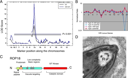
Genetic mapping of ROP18 as a major virulence determinant in the mouse. (A) Whole-genome analyses of the traits of acute mortality (vir) and serum responsiveness (serum) show a strong peak on chromosome VIIa with a minor peak on chromosome Ia. The x axis shows the 175 informative markers aligned across the 14 chromosomes. LOD score, log odds ratio. (B) Microarray analysis of a cluster of 21 genes from the central region of the QTL shown in panel A. Wild-type strains are untagged strains of the type I (GT-1) and type III (CTG) backgrounds, while “parental” refers to the drug-resistant strains used in the cross. (Reproduced from reference .) (C) Schematic of ROP18 showing the signal peptide, processing site, N-terminal domain containing low-complexity regions, and S/T kinase domain. (D) Discharge of rhoptries accompanies cell invasion. An empty rhoptry profile of a recently invaded tachyzoite (R) is visible. Bar, 200 nm.
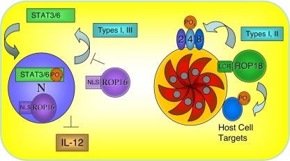
Model for the proposed roles of ROP16 and ROP18. (Right) ROP18 is highly expressed in type I and II strains, and following secretion it is targeted to the PVM by a series of low-complexity helical regions (LCR) in the N terminus. ROP18 has been shown to phosphorylate other ROPs in vitro, suggesting that it may also perform this function in infected cells (i.e., ROP2, -4, and -8, as shown). Additionally, host cell proteins may be targets of ROP18. The virulence-enhancing potential of ROP18 requires kinase activity and has been associated with slightly enhanced growth. (Left) ROP16 is targeted to the host cell nucleus (N) by a nuclear localization sequence (NLS), although it may also be active in the cytosol. ROP16 from type I and III strains induces prolonged activation of STAT3/6, thus activating gene expression in the host cell nucleus but blocking the production of IL-12.
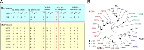
Divergence of ROP2 family members and degeneracy in the conserved kinase domains. (A) Alignment of several mammalian kinases (PKA-Cα and Jun N-terminal protein kinase [JNK]) showing conservation of key residues implicated in binding to ATP and in catalysis. The catalytic triad is boxed in red. While a majority of ROP2 family members are divergent and are predicted to be inactive, ROP16, ROP17, and ROP18 conserve the key residues associated with activity. (B) Phylogenetic tree showing the relationship between ROP kinases, FIKK kinases from P. falciparum, and major families of human kinases. TK, tyrosine kinases; AGC, cAMP-regulated kinase, c-GMP regulated kinase, and PKC; CAMK, calcium-regulated kinases; GMCC, cyclin-dependent, mitogen-activated, and casein kinases. (Reproduced from reference .)
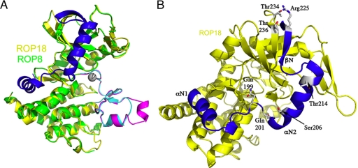
Ribbon models of the ROP8 and ROP18 kinase domains. (A) Model of ROP18 derived by homology modeling (yellow), superimposed on a model derived from the X-ray crystal structure of ROP8 (green). The N-terminal extension of the kinase domain is found in both proteins and is shown in blue. This extension consists of a β-sheet that completes the N lobe and two alpha helixes that wrap the main kinase domain. Substantial differences also occur in the substrate-binding domain, where ROP18 is shown in cyan and ROP8 in magenta. (B) Model of ROP18 showing residues that are phosphorylated and implicated in the regulation of activity. (Reproduced from reference .)
Similar articles
-
The Toxoplasma pseudokinase ROP5 forms complexes with ROP18 and ROP17 kinases that synergize to control acute virulence in mice.Cell Host Microbe. 2014 May 14;15(5):537-50. doi: 10.1016/j.chom.2014.04.002. Cell Host Microbe. 2014. PMID: 24832449 Free PMC article.
-
The Toxoplasma pseudokinase ROP5 is an allosteric inhibitor of the immunity-related GTPases.J Biol Chem. 2014 Oct 3;289(40):27849-58. doi: 10.1074/jbc.M114.567057. Epub 2014 Aug 12. J Biol Chem. 2014. PMID: 25118287 Free PMC article.
-
The Toxoplasma protein ARO mediates the apical positioning of rhoptry organelles, a prerequisite for host cell invasion.Cell Host Microbe. 2013 Mar 13;13(3):289-301. doi: 10.1016/j.chom.2013.02.001. Cell Host Microbe. 2013. PMID: 23498954
-
Genetic Mapping of Pathogenesis Determinants in Toxoplasma gondii.Annu Rev Microbiol. 2016 Sep 8;70:63-81. doi: 10.1146/annurev-micro-091014-104353. Epub 2016 Jun 17. Annu Rev Microbiol. 2016. PMID: 27359216 Review.
-
[Toxoplasma gondii proteome].Wiad Parazytol. 2003;49(1):3-10. Wiad Parazytol. 2003. PMID: 16889012 Review. Polish.
Cited by
-
A major locus confers triclabendazole resistance in Fasciola hepatica and shows dominant inheritance.PLoS Pathog. 2023 Jan 26;19(1):e1011081. doi: 10.1371/journal.ppat.1011081. eCollection 2023 Jan. PLoS Pathog. 2023. PMID: 36701396 Free PMC article.
-
Genomics of apicomplexan parasites.Crit Rev Biochem Mol Biol. 2017 Jun;52(3):254-273. doi: 10.1080/10409238.2017.1290043. Epub 2017 Feb 22. Crit Rev Biochem Mol Biol. 2017. PMID: 28276701 Free PMC article. Review.
-
Genetic Crosses and Linkage Mapping in Schistosome Parasites.Trends Parasitol. 2018 Nov;34(11):982-996. doi: 10.1016/j.pt.2018.08.001. Epub 2018 Aug 24. Trends Parasitol. 2018. PMID: 30150002 Free PMC article. Review.
-
Isolation and Proteomic Analysis of Rhoptry-Enriched Fractions from Cryptosporidium parvum.Iran J Public Health. 2015 Sep;44(9):1187-95. Iran J Public Health. 2015. PMID: 26587492 Free PMC article.
-
Modulation of innate immunity by Toxoplasma gondii virulence effectors.Nat Rev Microbiol. 2012 Nov;10(11):766-78. doi: 10.1038/nrmicro2858. Nat Rev Microbiol. 2012. PMID: 23070557 Free PMC article. Review.
References
-
- Ajioka, J. W., and L. D. Sibley. 2007. Development and application of classical genetics in Toxoplasma gondii, p. 367-389. In L. M. Weiss and K. Kim (ed.), Toxoplasma gondii the model Apicomplexan: perspectives and methods. Elsevier/Academic Press, New York, NY.
-
- Barragan, A., and L. D. Sibley. 2003. Migration of Toxoplasma gondii across biological barriers. Trends Microbiol. 11426-430. - PubMed
-
- Beckers, C. J. M., J. F. Dubremetz, O. Mercereau-Puijalon, and K. A. Joiner. 1994. The Toxoplasma gondii rhoptry protein ROP2 is inserted into the parasitophorous vacuole membrane, surrounding the intracellular parasite, and is exposed to the host cell cytoplasm. J. Cell Biol. 127947-961. - PMC - PubMed
-
- Boudeau, J., D. Miranda-Saavedra, G. J. Barton, and D. R. Alessi. 2006. Emerging roles of pseudokinases. Trends Cell Biol. 16443-452. - PubMed
Publication types
MeSH terms
Substances
Grants and funding
LinkOut - more resources
Full Text Sources
Medical
