The single substitution I259T, conserved in the plasminogen activator Pla of pandemic Yersinia pestis branches, enhances fibrinolytic activity
- PMID: 19465664
- PMCID: PMC2715710
- DOI: 10.1128/JB.00489-09
The single substitution I259T, conserved in the plasminogen activator Pla of pandemic Yersinia pestis branches, enhances fibrinolytic activity
Abstract
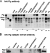
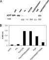
The outer membrane plasminogen activator Pla of Yersinia pestis is a central virulence factor in plague. The primary structure of the Pla beta-barrel is conserved in Y. pestis biovars Antiqua, Medievalis, and Orientalis, which are associated with pandemics of plague. The Pla molecule of the ancestral Y. pestis lineages Microtus and Angola carries the single amino acid change T259I located in surface loop 5 of the beta-barrel. Recombinant Y. pestis KIM D34 or Escherichia coli XL1 expressing Pla T259I was impaired in fibrinolysis and in plasminogen activation. Lack of detectable generation of the catalytic light chain of plasmin and inactivation of plasmin enzymatic activity by the Pla T259I construct indicated that Microtus Pla cleaved the plasminogen molecule more unspecifically than did common Pla. The isoform pattern of the Pla T259I molecule was different from that of the common Pla molecule. Microtus Pla was more efficient than wild-type Pla in alpha(2)-antiplasmin inactivation. Pla of Y. pestis and PgtE of Salmonella enterica have evolved from the same omptin ancestor, and their comparison showed that PgtE was poor in plasminogen activation but exhibited efficient antiprotease inactivation. The substitution (259)IIDKT/TIDKN in PgtE, constructed to mimic the L5 region in Pla, altered proteolysis in favor of plasmin formation, whereas the reverse substitution (259)TIDKN/IIDKT in Pla altered proteolysis in favor of alpha(2)-antiplasmin inactivation. The results suggest that Microtus Pla represents an ancestral form of Pla that has evolved into a more efficient plasminogen activator in the pandemic Y. pestis lineages.
Figures


Similar articles
-
Protein regions important for plasminogen activation and inactivation of alpha2-antiplasmin in the surface protease Pla of Yersinia pestis.Mol Microbiol. 2001 Jun;40(5):1097-111. doi: 10.1046/j.1365-2958.2001.02451.x. Mol Microbiol. 2001. PMID: 11401715
-
Lack of O-antigen is essential for plasminogen activation by Yersinia pestis and Salmonella enterica.Mol Microbiol. 2004 Jan;51(1):215-25. doi: 10.1046/j.1365-2958.2003.03817.x. Mol Microbiol. 2004. PMID: 14651623
-
Fibrinolytic and procoagulant activities of Yersinia pestis and Salmonella enterica.J Thromb Haemost. 2015 Jun;13 Suppl 1:S115-20. doi: 10.1111/jth.12932. J Thromb Haemost. 2015. PMID: 26149012 Review.
-
Yersinia pestis Plasminogen Activator.Biomolecules. 2020 Nov 14;10(11):1554. doi: 10.3390/biom10111554. Biomolecules. 2020. PMID: 33202679 Free PMC article. Review.
-
Using every trick in the book: the Pla surface protease of Yersinia pestis.Adv Exp Med Biol. 2007;603:268-78. doi: 10.1007/978-0-387-72124-8_24. Adv Exp Med Biol. 2007. PMID: 17966423 Review.
Cited by
-
Fibrinolytic and coagulative activities of Yersinia pestis.Front Cell Infect Microbiol. 2013 Jul 26;3:35. doi: 10.3389/fcimb.2013.00035. eCollection 2013. Front Cell Infect Microbiol. 2013. PMID: 23898467 Free PMC article. Review.
-
Cpa, the outer membrane protease of Cronobacter sakazakii, activates plasminogen and mediates resistance to serum bactericidal activity.Infect Immun. 2011 Apr;79(4):1578-87. doi: 10.1128/IAI.01165-10. Epub 2011 Jan 18. Infect Immun. 2011. PMID: 21245266 Free PMC article.
-
Phylogenetically Defined Isoforms of Listeria monocytogenes Invasion Factor InlB Differently Activate Intracellular Signaling Pathways and Interact with the Receptor gC1q-R.Int J Mol Sci. 2019 Aug 24;20(17):4138. doi: 10.3390/ijms20174138. Int J Mol Sci. 2019. PMID: 31450632 Free PMC article.
-
Yersinia virulence factors - a sophisticated arsenal for combating host defences.F1000Res. 2016 Jun 14;5:F1000 Faculty Rev-1370. doi: 10.12688/f1000research.8466.1. eCollection 2016. F1000Res. 2016. PMID: 27347390 Free PMC article. Review.
-
Early emergence of Yersinia pestis as a severe respiratory pathogen.Nat Commun. 2015 Jun 30;6:7487. doi: 10.1038/ncomms8487. Nat Commun. 2015. PMID: 26123398 Free PMC article.
References
-
- Achtman, M., G. Morelli, P. Zhu, T. Wirth, I. Diehl, B. Kusecek, A. J. Vogler, D. M. Wagner, C. J. Allender, W. R. Easterday, V. Chenal-Francisque, P. Worsham, N. R. Thomson, J. Parkhill, L. E. Lindler, E. Carniel, and P. Keim. 2004. Microevolution and history of the plague bacillus, Yersinia pestis. Proc. Natl. Acad. Sci. USA 10117837-17842. - PMC - PubMed
-
- Bergmann, S., and S. Hammerschmidt. 2007. Fibrinolysis and host response in bacterial infections. Thromb. Haemost. 98512-520. - PubMed
Publication types
MeSH terms
Substances
LinkOut - more resources
Full Text Sources

