. 2009 Jun;41(6):746-52.
doi: 10.1038/ng.380. Epub 2009 May 24.

Mutations in the beta-tubulin gene TUBB2B result in asymmetrical polymicrogyria

Affiliations

Mutations in the beta-tubulin gene TUBB2B result in asymmetrical polymicrogyria

Xavier Hubert Jaglin et al. Nat Genet. 2009 Jun.

Abstract

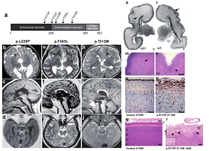
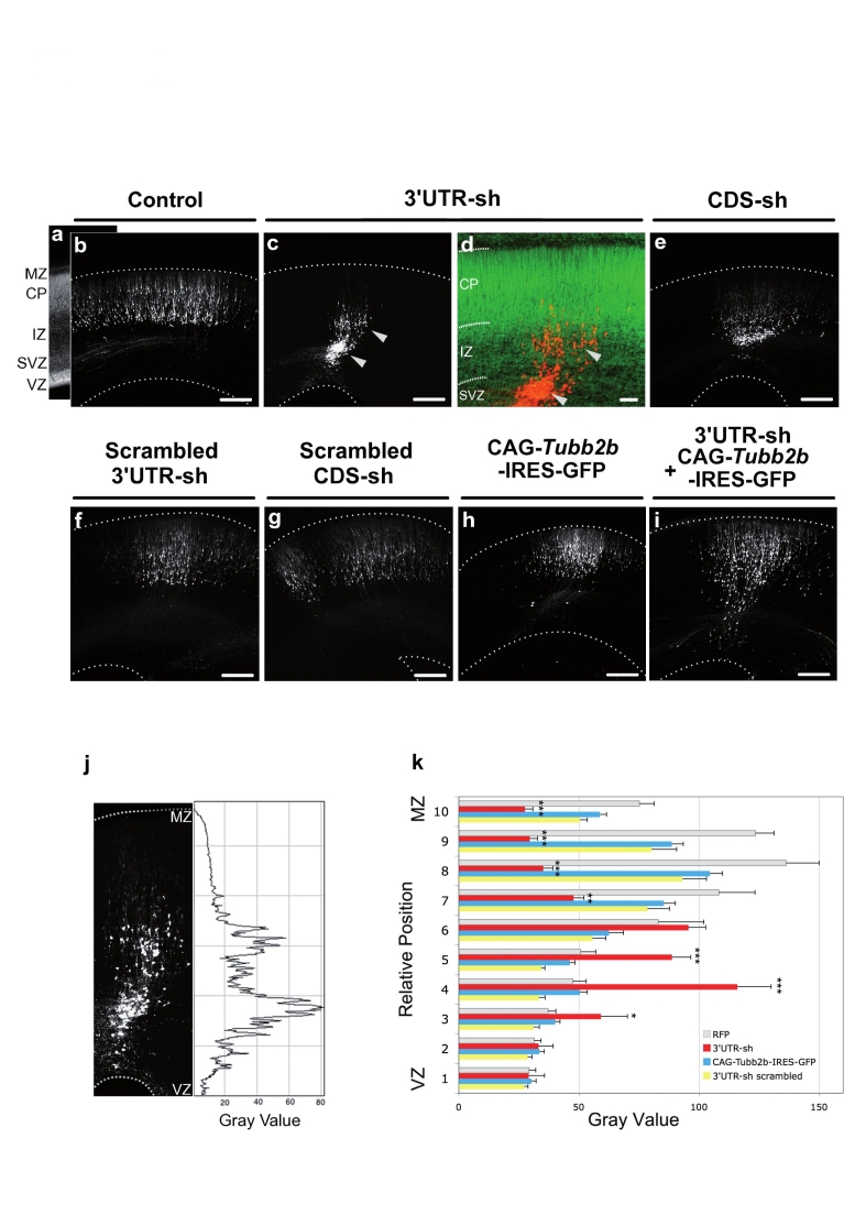
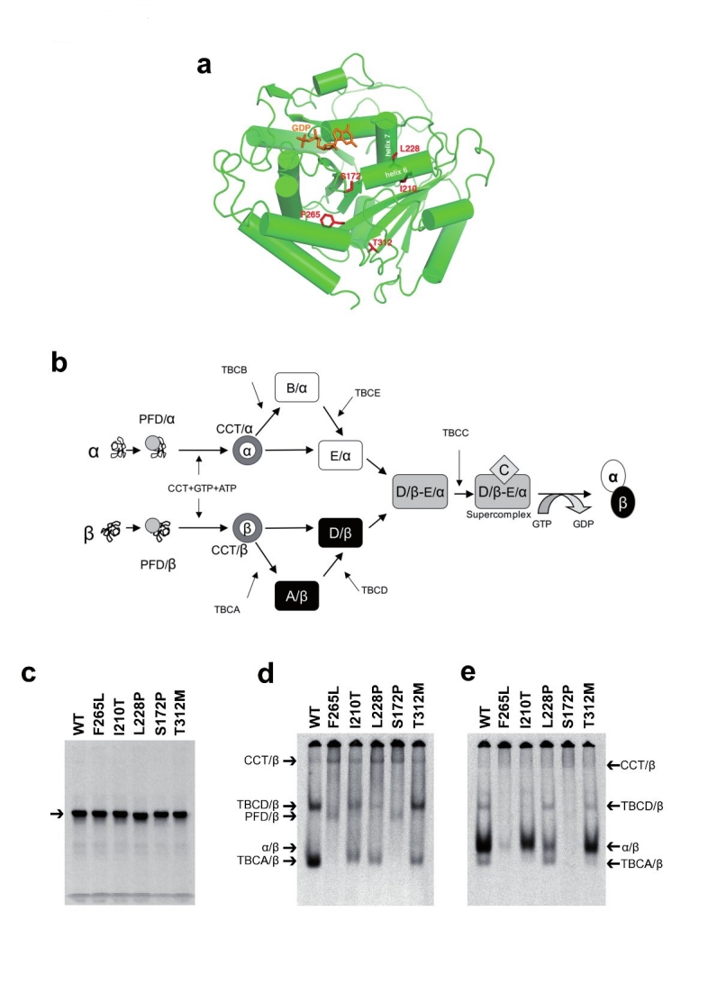
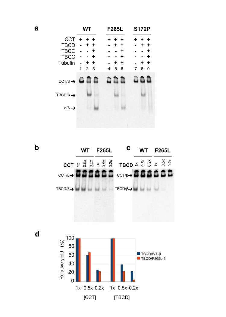
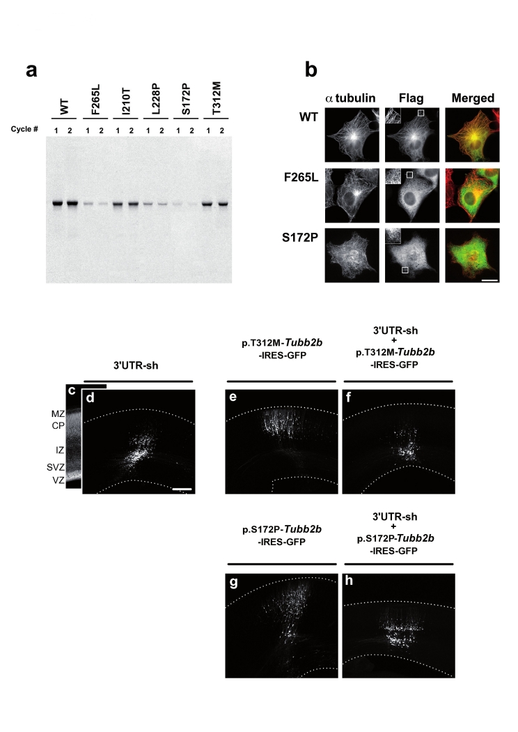
Polymicrogyria is a relatively common but poorly understood defect of cortical development characterized by numerous small gyri and a thick disorganized cortical plate lacking normal lamination. Here we report de novo mutations in a beta-tubulin gene, TUBB2B, in four individuals and a 27-gestational-week fetus with bilateral asymmetrical polymicrogyria. Neuropathological examination of the fetus revealed an absence of cortical lamination associated with the presence of ectopic neuronal cells in the white matter and in the leptomeningeal spaces due to breaches in the pial basement membrane. In utero RNAi-based inactivation demonstrates that TUBB2B is required for neuronal migration. We also show that two disease-associated mutations lead to impaired formation of tubulin heterodimers. These observations, together with previous data, show that disruption of microtubule-based processes underlies a large spectrum of neuronal migration disorders that includes not only lissencephaly and pachygyria, but also polymicrogyria malformations.


Figures

Figure 1
Figure 1. Magnetic Resonance Imaging and Histopathology Analysis of Patients with TUBB2B mutations
(a) Linear representation of the β-tubulin protein showing the position of heterozygous PMG-associated mutations. (b–j) Representative brain imaging features of 3 patients carrying TUBB2B mutations: P2 (p.L228P) (b-d); P3 (p.F265L) (e-g); P1 (p.T312M) (h-j). Axial images show areas of PMG that appear more severe in frontal and parietal lobes (b,e) and involve the perisylvian region (e,h). The PMG appears either mildly (b,h) or severely asymmetric with left-sided predominance (e) (Hatched lines highlighted by arrowheads show some of the PMG areas). Basal ganglia appear dysmorphic with a fusion of caudate and putamen, and apparent absence of the anterior arm of the internal capsule (b,e,h). Midline sagittal section shows corpus callosum agenesis (c), hypogenesis and abnormal thickness (f), or dysmorphy with a flat shape (i), associated with mild to severe cerebellar vermis hypoplasia or atrophy (c,f,i) and with brainstem hypoplasia (double asterisks). Axial section at the level of the cerebellum and temporal lobes show severe vermian dysplasia (d,j) or atrophy (g) (black asterisks). (k–n,q,r) Nissl-stained sections of the 27GW fetus (p.S172P mutation) brain display asymmetrical bilateral polymicrogyria (black arrows in k,l) with callosal agenesis. Left and right hemispheres present respectively typical unlayered polymicrogyric cortex (l,n) and focal polymicrogyria with a completely disorganized cortex and radial neuronal heterotopias (black arrows in k,m). Several nodular heterotopic neuron clusters were also observed in both hemispheres. Sections of cortical regions of left hemisphere probed for vimentin (o,p) or stained by Nissl (q,r) respectively show either abnormalities of radial glial fiber organization (p) or a disorganized cortex (r) with neuronal overmigration through the pial basement membrane into the leptomeningeal space (black arrows in r). Sections (o,q) correspond to a 27GW control fetus. M: meninges. Scale bars: (k–n) 500 μm, (o–r) 100 μm.
Figure 2
Figure 2. In Utero Knock-Down of rat Tubb2b Expression by RNAi Alters Neuronal Migration in the Isocortex
(a-i) Nissl staining on coronal sections of E20 brains reveals the overall organization of the cortex (a) 5 days after electroporation of a RFP coding reporter construct either alone (b) or in combination with 3′UTR-sh (c, overlay with Nissl in d), CDS-sh (e), or their corresponding scrambled controls (f,g). A rescue experiment was performed using CAG-Tubb2b-IRES-GFP transfected either alone (h, GFP), or combined with 3′UTR-sh (i, GFP). (j,k) Fluorescence intensities reflecting cell positions were converted into gray values and measured across cortices from the VZ to the MZ (j, 3′UTR-sh). k, Bars represent the mean ± SEM of fluorescence intensities in 10 strata dividing the thickness of cortices of independent brains (RFP n=5, 3′UTR-sh n=6, CDS-sh n=6, Scrambled 3′UTR-sh n=4, Scrambled CDS-sh n=5, Rescue n=6). Knockdown of Tubb2b using both hairpins between E15 and E20 disrupts neuronal migration (c,d,e). RFP positive cells are significantly stalled within the deep layers of the cortex (c,k, strata 4,5: F(3,42)>20.4, p<0.0001; p<0.001(***) for 3′UTR-sh compared by Tukey-Kramer test to RFP, scrambled 3′UTR-sh and Rescue respectively) that correspond to the SV/IZ (d, higher magnification) whereas neurons have already reached the CP in control conditions (b,f,g). Tubb2b overexpression preserves neuronal migration (h) showing that migration disruption is a specific consequence of Tubb2b RNAi as it rescued the defect (i,k strata 8-10: F(3,42)>23.7, p<0.0001, p<0.001(***) for 3′UTR-sh compared by Tukey-Kramer test to RFP and Rescue respectively). Hatched lines in (b,c,e-i) correspond to the outer/ventricular limits of the cortex. Scale bars : 200 µm (a-i).
Figure 3
Figure 3. Various Mutations in TUBB2B Result in Inefficient α/β Tubulin Heterodimer Formation In Vitro
(a) Ribbon presentation illustrating placement of side chains of mutated residues (shown in red) and the E-site guanine nucleotide (shown in orange) within the structure of the β-tubulin polypeptide[11,12].S172 resides between two proline residues in a loop. (b) The tubulin folding pathway involves a series of molecular chaperones whose function is to facilitate the assembly of the α/β tubulin heterodimer[25]. Newly translated α-tubulin (α) and β-tubulin (β) polypeptides are first captured and stabilized by prefoldin (PFD) that acts as a shuttling protein to deliver its bound target protein to the cytosolic chaperonin (CCT)[26]. CCT generates productive quasi-native folding intermediates which interact with a set of downstream Tubulin-specific Chaperones (TBCs)[27]. TBCB and TBCE capture CCT-generated α-tubulin intermediates in which the encapsulating GTP-binding pocket (the N-site) is already formed28 producing TBCB/α-tubulin (B/α) and TBCE/α-tubulin (E/α) cocomplexes. TBCA and TBCD capture and stabilize CCT-generated β-tubulin intermediates forming TBCA/β-tubulin (A/β) and TBCD/β-tubulin (D/β) cocomplexes. TBCD/β-tubulin (D/β) and TBCE/α-tubulin (E/α) converge to form a supercomplex with TBCC (C-D/β−E/α). Interaction with TBCC (C) results in the triggering of GTP hydrolysis by β-tubulin16. This reaction acts as a switch to signal the release of newly formed GDP-bound α/β heterodimers, which are then competent for incorporation into microtubules. (c-e) Analysis by SDS-PAGE (c) or non-denaturing gels (d,e) of in vitro transcription/translation products performed with wild-type (WT) and mutant TUBB2B and further chased with bovine brain tubulin so as to generate α/β tubulin heterodimers (e). The different migration pattern for p.L228P in (c) can be explained by the substitution of a proline in place of leucine, disrupting the helix, and presumably resulting in a change in the binding of SDS and hence a slight change in migration rate on the SDS gel. Note that p.F265L and p.S172P mutants yielded either only a trace or no discernable amount of α/β heterodimer. The remaining mutants all generated products present in the WT control, but in varying yield (d,e).
Figure 4
Figure 4. In Vitro Reactions Reveal a Lowered Affinity of p.S172P TUBB2B for TBCD
(a) Analysis on non-denaturing gels of the products of reconstituted folding reactions containing ATP, GTP and various combinations of the purified components that are essential to the heterodimer assembly reaction. (b–c) Analysis on non-denaturing gels of the products of in vitro folding reactions performed with 35S-methionine-labeled, unfolded wild type (WT) or p.F265L mutant protein. Reactions contained a range of concentrations of purified cytosolic chaperonin (CCT) (1x, 0.5x, 0.2x) in the presence of constant (1x molar equivalent) TBCD (control reaction) (b) or a range of concentrations of purified TBCD (1x, 0.5x, 0.2x) in the presence of constant (1x molar equivalent) CCT (c). (d) Quantitation of the data shown in (b) and (c). Note that when the abundance of TBCD was reduced by a factor of 5 in reconstituted reactions performed with a constant level of CCT, the yield of the TBCD/β-tubulin co-complex declined in the case of the wild type protein to 25% of the original level, but declined to an undetectable level in the case of p.F265L. Similar data were obtained in the case of the p.S172P mutation (Supplementary Fig. 8). The level of radioactivity present in complexes at the 1x concentration is taken as 100. Bars represent the average of two experiments. Arrows in (a–c) denote the migration positions of the CCT/β-tubulin binary complex, the TBCD/β-tubulin co-complex, the native tubulin heterodimer (α/β).
Figure 5
Figure 5. Loss of function of mutant TUBB2B in vitro, in cultured cells and in vivo.
(a) Copolymerization of labeled translation products with native bovine brain microtubules. Aliquots from two successive polymerization/depolymerization cycles (1 and 2), show inefficient incorporation for F265L, L228P and S172P mutants. (b) Expression of C-terminally FLAGtagged wild-type and mutant (p.S172P and p.F265L) TUBB2B in vivo after construct transfection into COS-7 cells. Note that F265L and S172P mutants do not incorporate into the MT network. (c-h) Nissl staining on coronal sections of E20 brains reveals the overall organization of the cortex (c), and repartition of GFP+ cells within the E20 cortices thickness, on coronal sections (d-h). Expression of p.T312M and p.S172P either alone (e,g) or in combination with 3′UTR-sh (f,h). Note that the over-expression of these mutants in combination with the hairpin does not rescue the neuronal migration defect caused by the RNAi (e,f) although the expression of each mutant alone does not lead to a major migration defect. However, we can see that upon expression of p.S172P a few cells seem to be blocked within the IZ suggesting that p.S172P could have a dominant effect leading to slight migration impairments. Scale bar: 200 µm.

Comment in

References

    1. Dutcher SK. The tubulin fraternity: alpha to eta. Curr Opin Cell Biol. 2001;13:49–54. - PubMed
    1. Keays D.A, et al. Mutations in alpha-tubulin cause abnormal neuronal migration in mice and lissencephaly in humans. Cell. 2007;128:45–57. - PMC - PubMed
    1. Poirier K, et al. Large spectrum of lissencephaly and pachygyria phenotypes resulting from de novo missense mutations in tubulin alpha 1A(TUBA1A). Hum Mutat. 2007;28:1055–64. - PubMed
    1. Bahi-Buisson N, et al. Refinement of cortical dysgeneses spectrum associated with TUBA1A mutations. J Med Genet. 2008;45:647–53. - PubMed
    1. Li S, et al. GPR56 regulates pial basement membrane integrity and cortical lamination. J Neurosci. 2008;28:5817–26. - PMC - PubMed

Publication types

MeSH terms

LinkOut - more resources