. 2009 Jun;11(6):739-46.
doi: 10.1038/ncb1880. Epub 2009 May 24.

The Patched dependence receptor triggers apoptosis through a DRAL-caspase-9 complex

Affiliations

The Patched dependence receptor triggers apoptosis through a DRAL-caspase-9 complex

Frédéric Mille et al. Nat Cell Biol. 2009 Jun.

Abstract

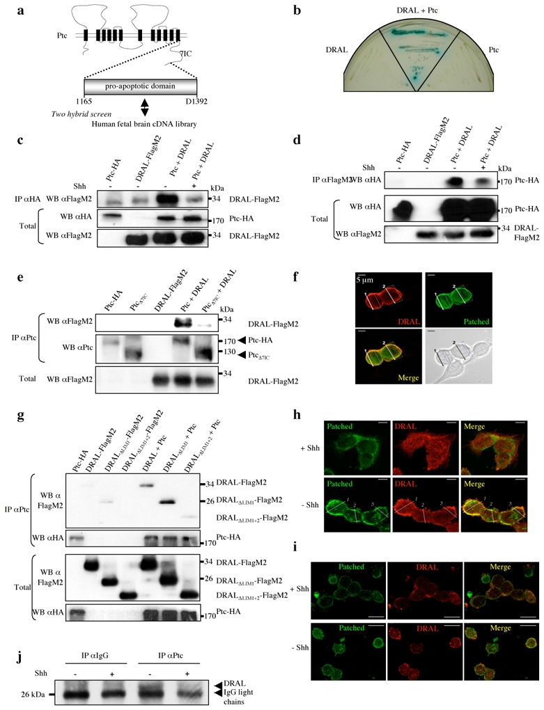
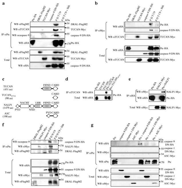
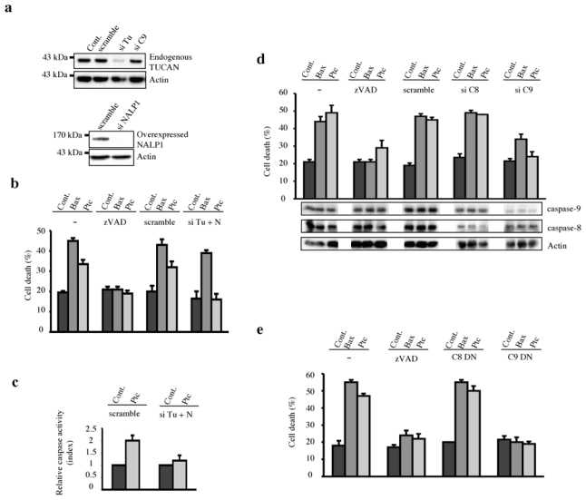
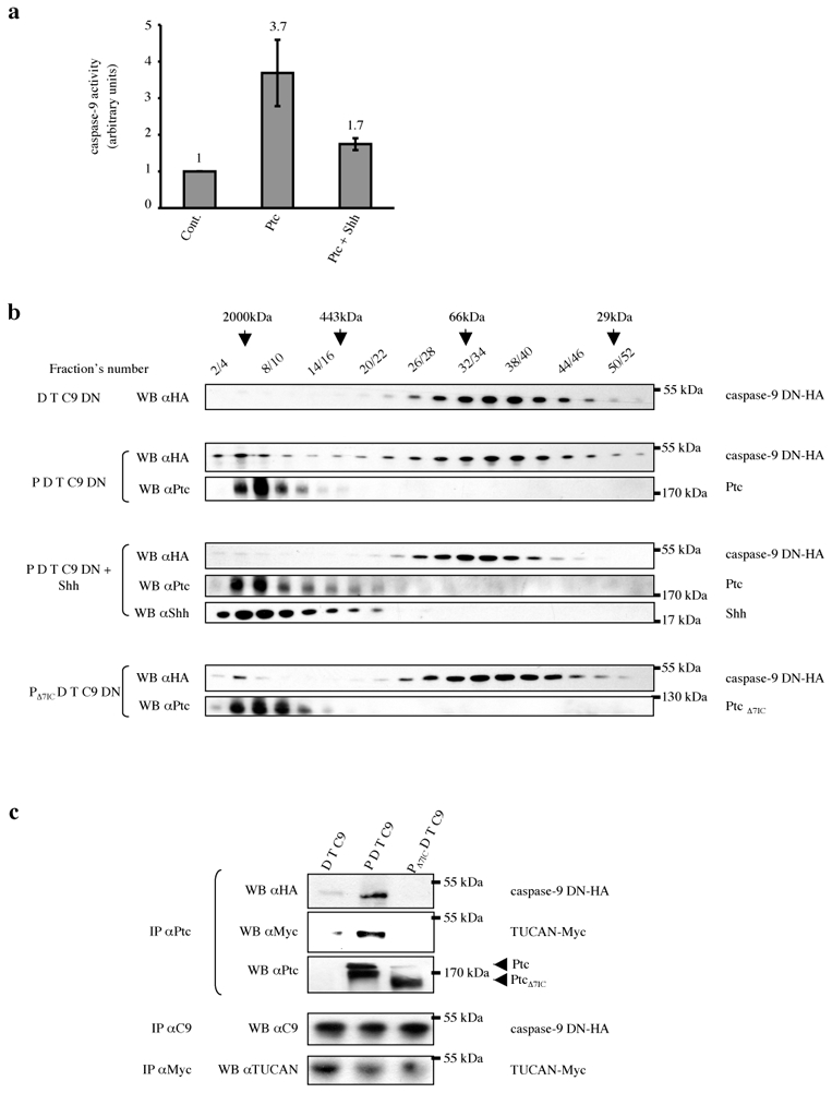
Sonic hedgehog (Shh) and its main receptor, Patched (Ptc), are implicated in both neural development and tumorigenesis. Besides its classic morphogenic activity, Shh is also a survival factor. Along this line, Ptc has been shown to function as a dependence receptor; it induces apoptosis in the absence of Shh, whereas its pro-apoptotic activity is blocked in the presence of Shh. Here we show that, in the absence of its ligand, Ptc interacts with the adaptor protein DRAL (downregulated in rhabdomyosarcoma LIM-domain protein; also known as FHL2). DRAL is required for the pro-apoptotic activity of Ptc both in immortalized cells and during neural tube development in chick embryos. We demonstrate that, in the absence of Shh, Ptc recruits a protein complex that includes DRAL, one of the caspase recruitment (CARD)-domain containing proteins TUCAN (family member, 8) or NALP1 (NLR family, pyrin domain containing 1) and apical caspase-9. Ptc triggers caspase-9 activation and enhances cell death through a caspase-9-dependent mechanism. Thus, we propose that in the absence of its ligand Shh the dependence receptor Ptc serves as the anchor for a caspase-activating complex that includes DRAL, and caspase-9.


Figures

Figure 1
Figure 1. The pro-apoptotic domain of Ptc interacts with DRAL
(a) A two-hybrid screen with the pro-apoptotic domain of Ptc-1. (b) The Ptc-DRAL interaction was confirmed by direct two-hybrid. AH109 yeasts transformed with either a mock Gal4BD plasmid and a Gal4AD plasmid fused with DRAL (DRAL) or a mock Gal4AD plasmid and a Gal4BD plasmid fused with the Ptc pro-apoptotic domain (Ptc). (c) Co-immunoprecipitations were performed on HEK293T cells transiently expressing Ptc-HA (Ptc) or DRAL-FlagM2 (DRAL) or Ptc-HA and DRAL-FlagM2 (Ptc + DRAL) in the absence (−) or presence (+) of Shh (300 ng/ml). Pull-down with anti-HA antibody was used to immunoprecipitate Ptc (IP αHA) and DRAL was revealed by Western blot by using anti-FlagM2 antibody. Western blot on lysates before pull down are shown (Total). (d) Same experiment as in (c) but in the opposite direction: DRAL pull-down was done with anti-FlagM2 antibody (IP αFlagM2) and specific Ptc binding was revealed by using anti-HA antibody. Full blots are shown in Suppl. Figure 7a. (e) DRAL co-immunoprecipitation with Ptc but not with PtcΔ7IC. Pull-down of Ptc on lysates from cells expressing DRAL-FlagM2 (DRAL) and Ptc-HA (Ptc) or PtcΔ7IC (PtcΔ7IC). DRAL was revealed by Western blot by using anti-FlagM2 antibody. (f) Co-localization of Ptc and DRAL in transiently transfected HEK293T cells. Cells were stained with anti-Ptc (Patched, green) and anti-DRAL (DRAL, red) antibodies. A representative field is presented from 3 independent experiments. Superimposed photograph (Merge) is shown. (g) Co-immunoprecipitation of Ptc with DRAL deletion mutants was performed as described in (c) on cells expressing Ptc-HA and DRAL-FlagM2, DRALΔLim1-FlagM2, or DRALΔLIM1+2-FlagM2. DRAL presence was revealed with anti-FlagM2 antibody. (h,i) Co-localization of endogenous Ptc and DRAL in Daoy medulloblastoma cells (h) or in primary cultured cerebellar granule neurons (i) performed in the presence or absence of Shh. Cells were stained as described in (f). Superimposed photographs (Merge) are shown. (j) Immunoprecipitation on mice cortex of E16.5 embryos incubated in absence (−) or presence (+) of Shh was performed using Ptc antibody (IP α-Ptc) or IgG as control (IP α-IgG), and DRAL was detected by immunoblot by using anti-DRAL antibody.
Figure 2
Figure 2. DRAL is required for Ptc pro-apoptotic activity
(a) HEK293T cells were transiently transfected with empty vector, siRNA scramble (scramble) or siRNA DRAL (si DRAL). 48 hours after siRNA transfection, cells were transfected a second time with empty vector (Cont.), Bax encoding vector (Bax) or Ptc encoding vector (Ptc) in the absence (−) or presence of zVAD-fmk (zVAD). Cell death was analyzed 24 hours after the second transfection by using trypan blue exclusion as described in . Standard deviations are indicated. siRNA DRAL efficiency on endogenous DRAL expression is shown by Western blotting using anti-DRAL antibody. Anti-GAPDH immunoblot is shown as a control of specificity and loading. (b) DRAL function in Ptc-induced apoptosis was also addressed in vivo in a context of absence of Shh created by removal of the caudal part of the Hensen node by microsurgery at E1.5 in the chick embryo in ovo. Before the microsurgery, the left side of the neural tube was electroporated with GFP and shDRAL-1 or scramble. Electroporation efficiency is shown on the upper picture (GFP) and TUNEL staining of the same section is shown on the lower picture (TUNEL). Scale bar : 100 μm. (c) Index of TUNEL positive cells is represented as the ratio between the number of cells stained in shDRAL-1 condition (shDRAL-1-electroporated side) to the number of cells stained in the control condition (Non-electroporated side) counted over 15 sections. Error bars indicate SEM. U test was performed (p=0.008). Note that DRAL shRNA electroporation alone or forced expression of DRAL had no significant effect on cell death (Suppl. Fig. 2ef).
Figure 3
Figure 3. Ptc/DRAL serves as a platform to recruit TUCAN or NALP1 and caspase-9
(a) Ptc pull-down (IP αPtc) on HEK293-EcrShh cells transfected with either Ptc (Ptc- HA), DRAL (DRAL-FlagM2), TUCAN (TUCAN-Myc) or caspase-9 DN (caspase-9 DN-HA) alone or the four constructs together in the absence (P D T C9) or presence (P D T C9 + Shh) of Shh (induction with Muristerone A plus recombinant Shh). DRAL, TUCAN and caspase-9 interaction with Ptc were revealed by Western blot by using anti-FlagM2, anti-Myc and anti-caspase-9 antibodies respectively. Full blots are shown in Suppl. Figure 7b. (b) TUCAN immunoprecipitation (IP αMyc) from HEK293T cells transfected with Ptc-HA, DRAL-FlagM2, TUCAN-Myc or caspase-9 DN-HA alone or the four constructs together in the absence (P D T C9) or presence (P D T C9 + Shh) of recombinant Shh; Ptc and caspase-9 were revealed after the pull-down by Western blotting using anti-HA antibody; TUCAN was revealed with anti-TUCAN antibody. Total: Western blots on lysate before pull-down. (c) Schematic representations of TUCAN, TUCAN mutant deleted of its N-Terminal domain, NALP1 and ASC. Note the domain of homology FIIND between TUCAN and NALP1 in their N-Terminal part (grey box). (d) TUCAN immunoprecipitation (using anti-TUCAN with an epitope in the CARD domain) from HEK293T cells transfected with empty vector and Ptc (Ptc-HA), TUCAN (TUCAN) or TUCAN deleted of its N-terminal part vectors (TUCANΔNter) or transfected with Ptc and either TUCAN (Ptc + TUCAN) or TUCAN deleted of its N-terminal region (Ptc + TUCANΔNter). Ptc was revealed in the pull-down by Western blot by using anti-HA antibody. (e) Ptc pull-down (IP αHA) on HEK293T cells transfected with empty vector and Ptc (Ptc-HA) or NALP1 vectors (NALP1-Myc) or transfected with Ptc and NALP1 (Ptc + NALP1). NALP1 pull-down with Ptc was revealed by anti-Myc Western blot. (f) Co-immunoprecipitations as in (a) except that TUCAN vector was replaced by NALP1 vector (NALP1-Myc). (g) Ptc pull-down (IP αPtc) on HEK293T cells transfected with Ptc vector (Ptc), caspase-9 DN (caspase-9 DN-HA), caspase-1 DN (caspase-1 DN-Myc) or ASC (ASC-Myc) alone or in combination with Ptc (Ptc + caspase-9 DN-HA; Ptc + caspase-1 DN-Myc; Ptc + ASC-Myc). Caspase-9, caspase-1 or ASC within the Ptc pull-down were detected using anti-HA or anti-Myc antibodies. Full blots are shown in Suppl. Figure 7c.
Figure 4
Figure 4. TUCAN/NALP1 and caspase-9 are required for Ptc-induced apoptosis
(a) siRNA TUCAN efficiency and specificity is shown by TUCAN Western blotting on endogenous TUCAN protein of HEK293T cells treated with scramble siRNA (scramble), siRNA TUCAN (si Tu) or siRNA caspase-9 (si C9). The same was done with NALP1 but siRNA NALP1 efficiency was assessed on HEK293T cells overexpressing NALP1. Anti-Actin immunoblot is shown as a control of specificity and loading. Full scanned blot is shown in Suppl. Figure 7d. (b) HEK293T cells were transfected with empty vector (Cont.), Bax or Ptc either without treatment (−) or in the presence of zVAD-fmk (zVAD), siRNA scramble (scramble), siRNA TUCAN plus siRNA NALP1 (si Tu + N) and cell death was analyzed by trypan blue exclusion assay. (c) Ptc-induced caspase activation is inhibited by a combination of siRNA TUCAN plus siRNA NALP1 as measured by relative caspase-3 activity. HEK293T cells were transfected with mock vector pcDNA3 (Cont.) or with Ptc in the presence of siRNA scramble (scramble) or siRNA TUCAN plus siRNA NALP1 (si Tu + N). Index of relative caspase activity is presented as the ratio between the caspase activity of the sample and that measured in HEK293T cells transfected with pcDNA3. Cell death was also compared in primary cultured cerebellar granule neurons from either CBA/J or C57Bl/6/J genetic background when deprived in Shh. While primary neurons death occurred in the CBA/J culture, no induction of cell death was detected in the C57Bl/6/J culture (Suppl. Fig.3). (d) HEK293T cells were transfected with empty vector (Cont.), Bax or Ptc either without treatment (−) or in the presence of zVAD-fmk (zVAD), siRNA scramble (scramble), siRNA caspase-8 (si C8) or siRNA caspase-9 (si C9) and cell death was analyzed by trypan blue exclusion assay. siRNA caspase-9 and caspase-8 efficiencies are shown by Western blotting using anti-caspase- 9 and anti-caspase-8 antibodies. Anti-Actin Western blot is shown as a control of specificity and loading. (e) HEK293T cells were co-transfected with empty vector (Cont.), Bax or Ptc with either empty vector (−), caspase-8 dominant-negative (C8 DN) or caspase-9 dominant-negative (C9 DN). Cell death was analyzed as in (d). For all experiments, error bars are s.d. n=3.
Figure 5
Figure 5. Ptc pro-apoptotic complex recruits and activates caspase-9
(a) HEK293-EcRShh cells were transfected with either empty vector (Cont.) or Ptc encoding vector in the absence (Ptc) or in the presence (Ptc + Shh) of Shh (induction with Muristerone A plus addition of 300 ng/ml of recombinant Shh). The cell lysates were subjected to caspase-9 activity assay over time using LEHD-AFC substrate of caspase-9. Error bars indicate s.d. n=4. (b) HEK293-EcRShh cells were transfected with DRAL-FlagM2, TUCAN-Myc and caspase-9 DN-HA vectors with either empty vector (D T C9) or Patched vector in the absence of Shh (P D T C9) or in the presence of Shh (P D T C9 + Shh; induction by Muristerone A plus recombinant Shh) or Ptc deleted of its last intracellular domain (PΔ7IC D T C9). Cell lysates were separated by gel filtration chromatography. Fractions were then analyzed by Western blotting for caspase-9 (WB αHA), Ptc (WB αPtc) and Shh (WB αShh). The fractions’ numbers and the fractions in which the molecular mass markers were eluted are shown. (c) The 20 first fractions were pooled and a pull-down of Ptc was performed (IP αPtc). Anti-HA and anti-Myc immunoblots were then performed and they show that caspase-9 and TUCAN interact with Ptc in these high molecular weight fractions. The relative quantity of proteins of interest (caspase-9 and TUCAN) in cell lysate of each condition was checked by immunoprecipitation (IP αC9 and IP αMyc respectively) and Western blotting with anti-caspase-9 and anti-TUCAN respectively.

References

    1. Jessell TM. Neuronal specification in the spinal cord: inductive signals and transcriptional codes. Nat Rev Genet. 2000;1:20–29. - PubMed
    1. Murone M, Rosenthal A, de Sauvage FJ. Sonic hedgehog signaling by the patched-smoothened receptor complex. Curr Biol. 1999;9:76–84. - PubMed
    1. Charrier JB, Lapointe F, Le Douarin NM, Teillet MA. Anti-apoptotic role of Sonic hedgehog protein at the early stages of nervous system organogenesis. Development. 2001;128:4011–4020. - PubMed
    1. Litingtung Y, Chiang C. Specification of ventral neuron types is mediated by an antagonistic interaction between Shh and Gli3. Nat Neurosci. 2000;3:979–985. - PubMed
    1. Thibert C, et al. Inhibition of neuroepithelial patched-induced apoptosis by sonic hedgehog. Science. 2003;301:843–846. - PubMed

Publication types

MeSH terms