Review
. 2009 Aug;66(8):618-34.
doi: 10.1002/cm.20385.

Cytoplasmic Ig-domain proteins: cytoskeletal regulators with a role in human disease

Affiliations
Review

Cytoplasmic Ig-domain proteins: cytoskeletal regulators with a role in human disease

Carol A Otey et al. Cell Motil Cytoskeleton. 2009 Aug.

Abstract

Immunoglobulin domains are found in a wide variety of functionally diverse transmembrane proteins, and also in a smaller number of cytoplasmic proteins. Members of this latter group are usually associated with the actin cytoskeleton, and most of them bind directly to either actin or myosin, or both. Recently, studies of inherited human disorders have identified disease-causing mutations in five cytoplasmic Ig-domain proteins: myosin-binding protein C, titin, myotilin, palladin, and myopalladin. Together with results obtained from cultured cells and mouse models, these clinical studies have yielded novel insights into the unexpected roles of Ig domain proteins in mechanotransduction and signaling to the nucleus. An emerging theme in this field is that cytoskeleton-associated Ig domain proteins are more than structural elements of the cell, and may have evolved to fill different needs in different cellular compartments. Cell Motil. Cytoskeleton 2009. (c) 2009 Wiley-Liss, Inc.


Figures

Figure 1
Figure 1. The crystal structure of telokin and the framework of an I-set Ig domain
The crystal structure of telokin (pdb code 1FHG), the founding member of the I-set, is shown from both sides as a ribbon representation of the backbone. Members of the I-set of Ig domains share a common fold pattern consisting of two sandwiched beta sheets, depicted as a two dimensional template (2b). The upper beta sheet consists of beta strands A, B, D, and E; the lower beta sheet consists of beta strands A’, C, C’, F, and G. The coloring of the strands in the template is the same as the coloring of the beta strands in the structures of telokin.
Figure 2
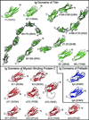
Figure 2. Structures of Ig domains from titin, palladin, and myosin binding protein-C
High-resolution structures of titin Ig domains have been solved for representative Ig domains from the proximal Ig segment (I1), the distal Ig segment (I27 and I65–I70), the P-zone of the A-band (A168–A169), the M-line (M1 and M5), and the Z-line (Z1–Z2). The A170 domain is a fibronectin type 3 (Fn3) domain that was solved in tandem with the A168 and A169 Ig domains (pdb code 2NZI). The palladin Ig3 and Ig4 domains are the only structures of Ig domains currently available from the palladin family of proteins. High-resolution structures are available for the fast-type isoform (fC1 and fC3), the cardiac isoform (cC1, cC2, and cC5), and the slow-type isoform (sC1, sC2, sC3, and sC4). The cC5 Ig domain contains an unstructured 28 amino acid insert between the C and D beta strands.
Figure 3
Figure 3. Location of Ig domains proteins within the sarcomere in striated muscle
MyBP-C is found in transverse stripes in the C-Zone. Myopalladin, palladin and myotilin localize to the Z line. Titin spans from the Z-line to the M-line.
Figure 4
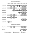
Figure 4. Myotilin-palladin-myopalladin family members
Schematic representation of myotilin-palladin-myopalladin family members and palladin’s major isoforms, and their domain organization. PR, proline-rich region; Ig, immunoglobulin domain.

References

    1. Ababou A, Rostkova E, Mistry S, Le Masurier C, Gautel M, Pfuhl M. Myosin binding protein C positioned to play a key role in regulation of muscle contraction: structure and interactions of domain C1. J Mol Biol. 2008;384:615–630. - PMC - PubMed
    1. Agarkova I, Ehler E, Lange S, Schoenauer R, Perriard JC. M-band: a safeguard for sarcomere stability? J Muscle Res Cell Motil. 2003;24:191–203. - PubMed
    1. Agarkova I, Perriard JC. The M-band: an elastic web that crosslinks thick filaments in the center of the sarcomere. Trends Cell Biol. 2005;15:477–485. - PubMed
    1. Aihara Y, Kurabayashi M, Saito Y, Ohyama Y, Tanaka T, Takeda S, Tomaru K, Sekiguchi K, Arai M, Nakamura T, Nagai R. Cardiac ankyrin repeat protein is a novel marker of cardiac hypertrophy: role of M-CAT element within the promoter. Hypertension. 2000;36:48–53. - PubMed
    1. Alyonycheva TN, Mikawa T, Reinach FC, Fischman DA. Isoform-specific interaction of the myosin-binding proteins (MyBPs) with skeletal and cardiac myosin is a property of the C-terminal immunoglobulin domain. J Biol Chem. 1997;272:20866–20872. - PubMed

Publication types

LinkOut - more resources