. 2009 May 25:6:62.
doi: 10.1186/1743-422X-6-62.

Hepatitis C virus NS4B carboxy terminal domain is a membrane binding domain

Affiliations

Hepatitis C virus NS4B carboxy terminal domain is a membrane binding domain

Jolanda M P Liefhebber et al. Virol J. .

Abstract

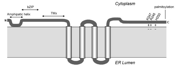
Background: Hepatitis C virus (HCV) induces membrane rearrangements during replication. All HCV proteins are associated to membranes, pointing out the importance of membranes for HCV. Non structural protein 4B (NS4B) has been reported to induce cellular membrane alterations like the membranous web. Four transmembrane segments in the middle of the protein anchor NS4B to membranes. An amphipatic helix at the amino-terminus attaches to membranes as well. The carboxy-terminal domain (CTD) of NS4B is highly conserved in Hepaciviruses, though its function remains unknown.

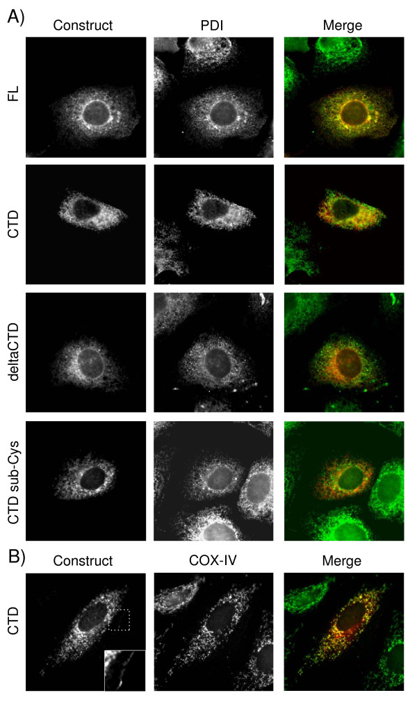
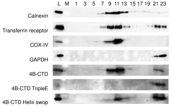
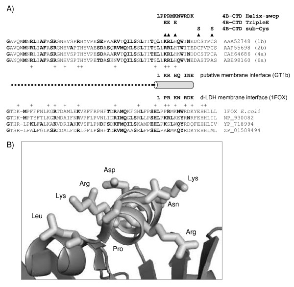
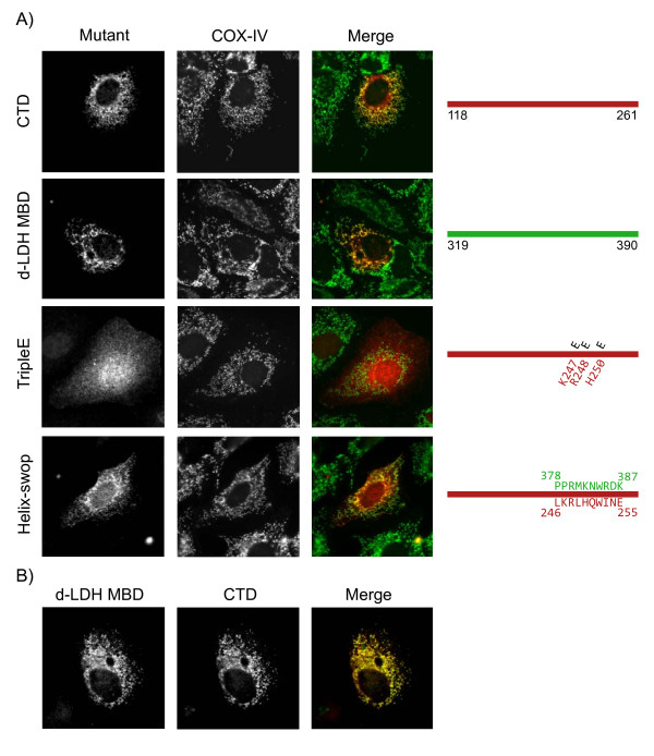
Results: A cytosolic localization is predicted for the NS4B-CTD. However, using membrane floatation assays and immunofluorescence, we now show targeting of the NS4B-CTD to membranes. Furthermore, a profile-profile search, with an HCV NS4B-CTD multiple sequence alignment, indicates sequence similarity to the membrane binding domain of prokaryotic D-lactate dehydrogenase (d-LDH). The crystal structure of E. coli d-LDH suggests that the region similar to NS4B-CTD is located in the membrane binding domain (MBD) of d-LDH, implying analogy in membrane association. Targeting of d-LDH to membranes occurs via electrostatic interactions of positive residues on the outside of the protein with negative head groups of lipids. To verify that anchorage of d-LDH MBD and NS4B-CTD is analogous, NS4B-CTD mutants were designed to disrupt these electrostatic interactions. Membrane association was confirmed by swopping the membrane contacting helix of d-LDH with the corresponding domain of the 4B-CTD. Furthermore, the functionality of these residues was tested in the HCV replicon system.

Conclusion: Together these data show that NS4B-CTD is associated to membranes, similar to the prokaryotic d-LDH MBD, and is important for replication.


Figures

Figure 1
Figure 1
Expression of different NS4B proteins in Huh7 cells. Huh7 cells were transfected with NS4B full-length (FL, aa 1–261), deltaCTD (aa 1–192), CTD (aa 188–261) or CTD substitution-Cysteines (CTD sub-Cys) and 24 h later processed for indirect immunofluorescence. Cells were double labeled with antibodies reacting against Myc-epitope-tag at the C-terminal end of each protein (in red) and A. protein disulphide isomerase (PDI) or B. Cytochrome C oxidase subunit IV (COX-IV) (in green), in first and second panels respectively. Third panels show merged images.
Figure 2
Figure 2
Membrane association of NS4B carboxy terminal domain. Huh7 cells transfected with NS4B-CTD-Myc, NS4B-CTD tripleE-Myc or NS4B-CTD Helix-swop-Myc were subjected to sucrose density gradient centrifugation. Cell lysates were loaded under a sucrose gradient from 10–80% w/v and part of the lysate was used as a loading control (L). Fractions were taken from top (fraction 1) to bottom (fraction 23) and separated by SDS-PAGE. Followed by immunoblot analysis for Calnexin, Transferrin Receptor (TfR), Cytochrome C oxidase subunit IV (COX-IV) and Glyceraldehyde 3-phosphate dehydrogenase (GAPDH). NS4B-CTD and NS4B-CTD tripleE were assayed using an antibody against Myc-epitope. M indicates where molecular weight marker was loaded.
Figure 3
Figure 3
Sequence similarity between NS4B carboxy terminal domain and the membrane binding domain of D-lactate dehydrogenase. A. Multiple sequence alignment of the carboxy terminal domain of four genotypes of HCV NS4B proteins and the membrane binding domain of four d-LDH family members (referenced by their accession numbers). Bold residues highlight amino-acids present in both families. Basic residues (R, K, H) making up the potential electropositive surface are indicated (+). Dotted line indicates disordered region in the d-LDH crystal structure. Arrowheads point to mutations made in the CTD of NS4B. B. Ribbon representation of the membrane anchored side of d-LDH (PDB code 1F0X). Stick residues indicate the surface exposed amino-acids of the ordered membrane binding helix.
Figure 4
Figure 4
Cellular distribution of NS4B carboxy terminal domain mutants and D-lactate dehydrogenase membrane binding domain in Huh7 cells. A. The panels on the right show Huh7 cells expressing different NS4B-CTD mutants or d-LDH membrane binding domain (d-LDH MBD) after 24 h. Expression constructs are shown in red. Using COX-IV as a marker protein, mitochondria are shown in middle panels and in merged picture in green. On the right a schematic view of the different NS4B-CTD mutants is drawn; in red the sequence of NS4B-CTD-wt, in black the mutations made and in green the exchanged amino acids from the membrane contacting helix of the d-LDH-MBD. B. Huh7 cells were co-transfected with NS4B-CTD-HA and d-LDH MBD-Myc and analyzed by immunofluorescence after 24 h of expression. The first panel shows d-LDH MBD, which is presented as red in the merged picture. NS4B-CTD is displayed in the second panel and is shown in the merged picture as green.
Figure 5
Figure 5
Effect of NS4B carboxy terminal mutations on colony formation using selectable replicons. Colony formation assay in which Huh7 cells are transfected with in vitro transcribed replicon RNA that contain NS4B-CTD mutations. Colonies were stained using Coomassie blue. Wild-type is pFK5.1. Mock transfected cells as the control. The NS4B-CTD mutations TripleE, E247, E248 and helix-swop in pFK5.1 are explained in figure 4.
Figure 6
Figure 6
Model of NS4B membrane association. Schematic of the proposed topology of NS4B relative to the ER membrane and reported functional properties (see introduction). Model for NS4B-CTD membrane association is discussed in this paper. Here we propose that positive residues (amino acids are indicated) are important for membrane targeting through the interaction with the negative head groups of phospholipids. Abbreviations: Nt, Amino terminus; Ct, Carboxyl terminus; bZIP, basic leucine zipper motif; TMx, transmembrane segment X.

References

    1. Gowans EJ. Distribution of markers of hepatitis C virus infection throughout the body. Semin Liver Dis. 2000;20:85–102. doi: 10.1055/s-2000-9503. - DOI - PubMed
    1. Hoofnagle JH. Course and outcome of hepatitis C. Hepatology. 2002;36:S21–S29. doi: 10.1002/hep.1840360704. - DOI - PubMed
    1. Lauer GM, Walker BD. Hepatitis C virus infection. N Engl J Med. 2001;345:41–52. doi: 10.1056/NEJM200107053450107. - DOI - PubMed
    1. Thiel HJ, Collett MS, Gould EA, Heinz FX, Houghton M, Meyers G. Virus Taxonomy: The Eighth Report of the International Committee on Taxonomy of Viruses (eds Fauquet, CM, Mayo, MA, Maniloff, J, Desselberger, U, and Ball, LA) 2005. pp. 979–996. Ref Type: Generic.
    1. van Leeuwen HC, Reusken CB, Roeten M, Dalebout TJ, Riezu-Boj JI, Ruiz J, Spaan WJ. Evolution of naturally occurring 5' non-translated region variants of hepatitis C virus genotype 1b in selectable replicons. J Gen Virol. 2004;85:1859–1866. doi: 10.1099/vir.0.79924-0. - DOI - PubMed

Publication types

Substances

LinkOut - more resources