Review
. 2009 May 26:11:e16.
doi: 10.1017/S1462399409001082.

Adhesion of Plasmodium falciparum-infected erythrocytes to human cells: molecular mechanisms and therapeutic implications

Affiliations
Free PMC article
Review

Adhesion of Plasmodium falciparum-infected erythrocytes to human cells: molecular mechanisms and therapeutic implications

J Alexandra Rowe et al. Expert Rev Mol Med. .
Free PMC article

Abstract

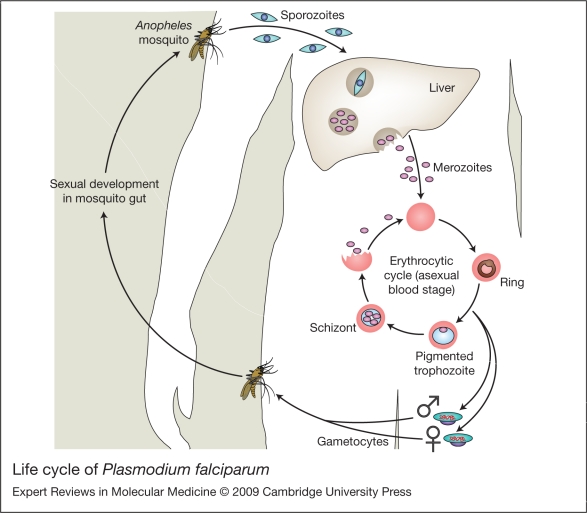
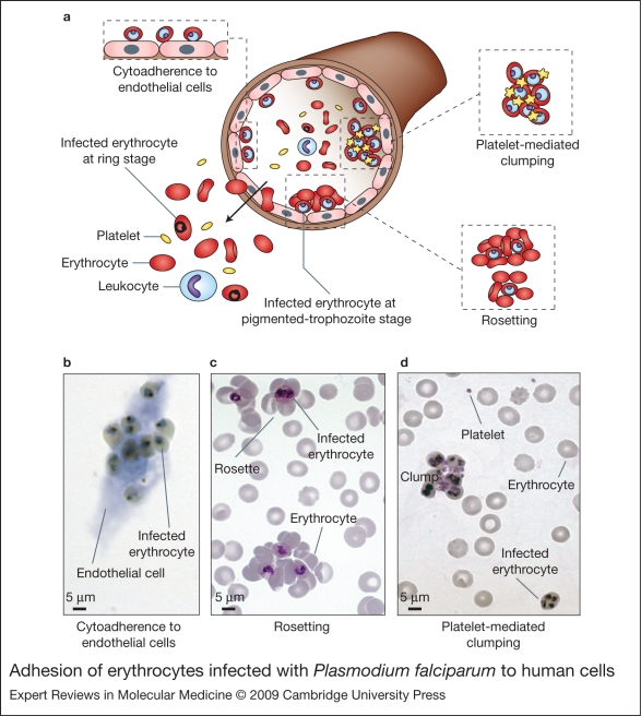
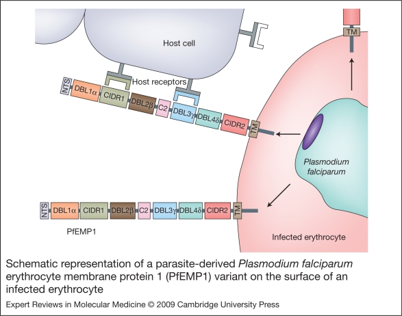
Severe malaria has a high mortality rate (15-20%) despite treatment with effective antimalarial drugs. Adjunctive therapies for severe malaria that target the underlying disease process are therefore urgently required. Adhesion of erythrocytes infected with Plasmodium falciparum to human cells has a key role in the pathogenesis of life-threatening malaria and could be targeted with antiadhesion therapy. Parasite adhesion interactions include binding to endothelial cells (cytoadherence), rosetting with uninfected erythrocytes and platelet-mediated clumping of infected erythrocytes. Recent research has started to define the molecular mechanisms of parasite adhesion, and antiadhesion therapies are being explored. However, many fundamental questions regarding the role of parasite adhesion in severe malaria remain unanswered. There is strong evidence that rosetting contributes to severe malaria in sub-Saharan Africa; however, the identity of other parasite adhesion phenotypes that are implicated in disease pathogenesis remains unclear. In addition, the possibility of geographic variation in adhesion phenotypes causing severe malaria, linked to differences in malaria transmission levels and host immunity, has been neglected. Further research is needed to realise the untapped potential of antiadhesion adjunctive therapies, which could revolutionize the treatment of severe malaria and reduce the high mortality rate of the disease.


Figures

Figure 1
Figure 1
Life cycle of Plasmodium falciparum. When an infected female Anopheles mosquito takes a blood meal, sporozoite forms of P. falciparum are injected into the human skin. The sporozoites migrate into the bloodstream and then invade liver cells. The parasite grows and divides within liver cells for 8–10 days, then daughter cells called merozoites are released from the liver into the bloodstream, where they rapidly invade erythrocytes. Merozoites subsequently develop into ring-stage, pigmented-trophozoite-stage and schizont-stage parasites within the infected erythrocyte. P. falciparum-infected erythrocytes express parasite-derived adhesion molecules on their surface, resulting in sequestration of pigmented-trophozoite and schizont stages in the microvasculature. The asexual intraerythrocytic cycle lasts for 48 hours, and is completed by the formation and release of new merozoites that will re-invade uninfected erythrocytes. It is during this asexual bloodstream cycle that the clinical symptoms of malaria (fever, chills, impaired consciousness, etc.) occur. During the asexual cycle, some of the parasite cells develop into male and female sexual stages called gametocytes that are taken up by feeding female mosquitoes. The gametocytes are fertilised and undergo further development in the mosquito, resulting in the presence of sporozoites in the mosquito salivary glands, ready to infect another human host.
Figure 2
Figure 2
Adhesion of Plasmodium falciparum-infected erythrocytes to human cells. (Legend; See previous page for figure) (a) Schematic representation of the adhesion properties of P. falciparum-infected erythrocytes to different host cells. Erythrocytes infected with mature forms of P. falciparum parasites (pigmented trophozoites and schizonts) have the ability to bind to a range of host cells, such as endothelium, uninfected erythrocytes (rosetting) and platelets (platelet-mediated clumping). The adhesion of infected erythrocytes to endothelial cells leads to their sequestration in the microvasculature of various organs and tissues such as heart, lung, brain, muscle and adipose tissue. As a result, only erythrocytes carrying young ring forms of the parasite are detected in human peripheral blood samples. Although cytoadherence and sequestration of mature infected erythrocytes in the microvasculature occur in all infections, several specific adhesive phenotypes have been associated with severe pathological outcomes of malaria, such as the formation of rosettes and the adhesion of infected erythrocytes to brain endothelium. Rosetting and platelet-mediated clumping are phenotypes that are displayed by some but not all P. falciparum isolates in vitro. In vivo, it is thought that the formation of rosettes and clumps will be accompanied by adhesion to endothelial cells and sequestration in the microcapillaries (Ref. 115). (b) Cytoadherence of infected erythrocytes to in-vitro-cultured brain endothelial cells, visualised by light microscopy after Giemsa staining. (c) Rosettes detected in in vitro P. falciparum cultures, observed after preparation of Giemsa-stained thin smears and light microscopy. (d) Platelet-mediated clumps of infected erythrocytes formed after in vitro co-incubation of parasite cultures with platelets, observed by Giemsa-stained thin smears and light microscopy.
Figure 3
Figure 3
Schematic representation of a parasite-derived Plasmodium falciparum erythrocyte membrane protein 1 (PfEMP1) variant on the surface of an infected erythrocyte. PfEMP1 is a family of proteins encoded by var genes that are transported and expressed on the surface of infected erythrocytes during the mature stages of the intraerythrocytic cycle (pigmented trophozoite and schizont). There are approximately 60 var genes per parasite genome, which encode 60 different variants of PfEMP1; however, only one particular variant of PfEMP1 is expressed per cell at any given time. Switching of var gene expression allows the parasite to modify the antigenic and functional properties of infected erythrocytes, thereby evading immunity and altering adhesion capabilities. The extracellular region of PfEMP1 has an N-terminal segment (NTS) followed by several cysteine-rich domains known as DBL (duffy-binding-like) and CIDR (cystein-rich interdomain regions) that can be classified into distinct types based upon sequence similarity. There are six DBL types, (α, β, γ, δ, ɛ and X) and three CIDR types (α, β and γ). The number, location and type of DBL and CIDR domains vary among PfEMP1 variants, and this variable domain composition and extensive sequence polymorphism is thought to provide great flexibility in binding properties. To date, the binding domains for several host receptors, such as CD36, complement receptor 1 and ICAM1, have been mapped to individual DBL and CIDR domains. This diagram shows a hypothetical model of a PfEMP1 variant. TM, transmembrane region.

Comment in

References

    1. Snow R.W.. et al. The global distribution of clinical episodes of Plasmodium falciparum malaria. Nature. 2005;434:214–217. - PMC - PubMed
    1. Marsh K.. et al. Indicators of life-threatening malaria in African children. New England Journal of Medicine. 1995;332:1399–1404. - PubMed
    1. Dondorp A.M.. et al. The relationship between age and the manifestations of and mortality associated with severe malaria. Clinical Infectious Diseases. 2008;47:151–157. - PubMed
    1. Miller L.H.. et al. The pathogenic basis of malaria. Nature. 2002;415:673–679. - PubMed
    1. Dondorp A.M.. et al. Direct in vivo assessment of microcirculatory dysfunction in severe falciparum malaria. Journal of Infectious Diseases. 2008;197:79–84. - PubMed

Further reading, resources and contacts

Books
    1. D.A. Warrell, H.M. Gilles. 4th Edition. Oxford University Press; 2002. eds ( ) Essential Malariology,
    2. This book provides a broad background to malariology.

    1. I.W. Sherman. ASM Press; 2005. ed. ( ) Molecular Approaches to Malaria,
    2. A good overview of current molecular approaches to malaria research.

Websites
    1. http://www.cdc.gov/Malaria/ http://www.cdc.gov/Malaria/
    2. For general information on malaria:

    1. http://www.who.int/topics/malaria/en/ http://www.who.int/topics/malaria/en/
    2. For information on malaria parasite biology, biochemistry and physiology see:

    1. http://sites.huji.ac.il/malaria/ http://sites.huji.ac.il/malaria/

Publication types