. 2009 Jul 15;24(11):3208-14.
doi: 10.1016/j.bios.2009.03.037. Epub 2009 Apr 2.

Integrating microfluidics and lensless imaging for point-of-care testing

Affiliations

Integrating microfluidics and lensless imaging for point-of-care testing

Sangjun Moon et al. Biosens Bioelectron. .

Abstract

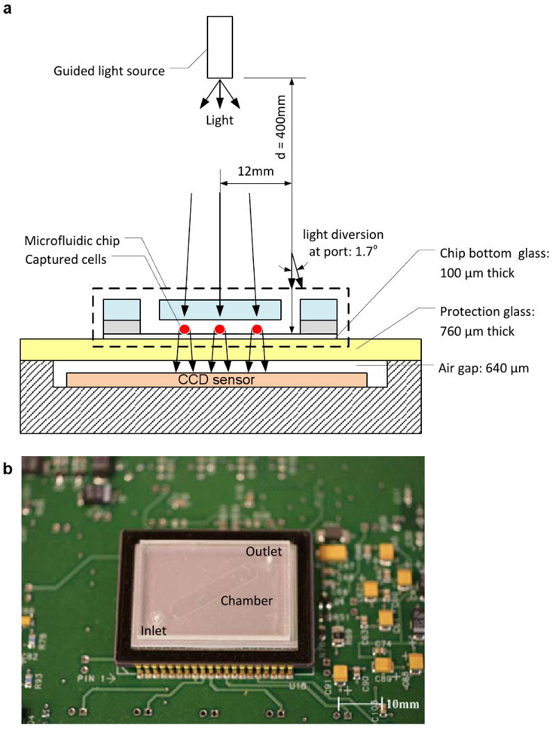
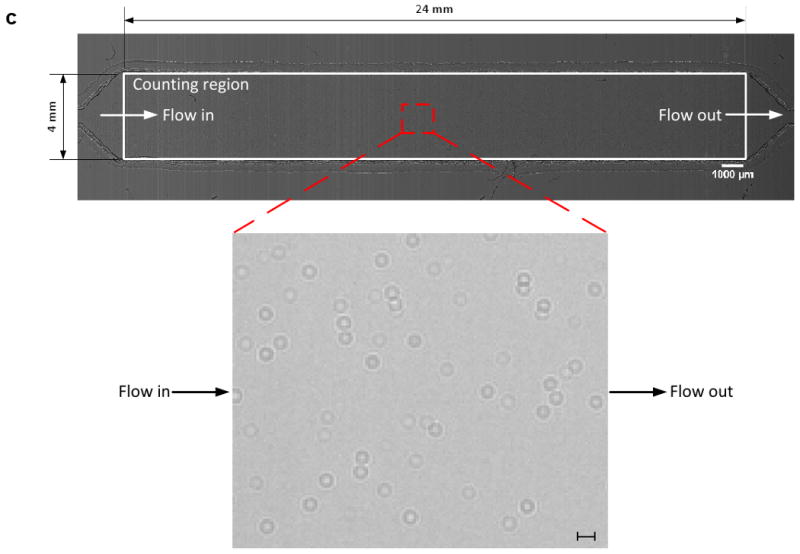
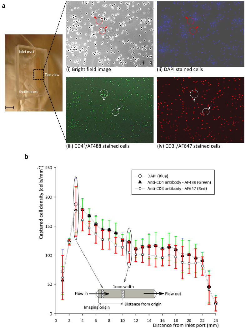
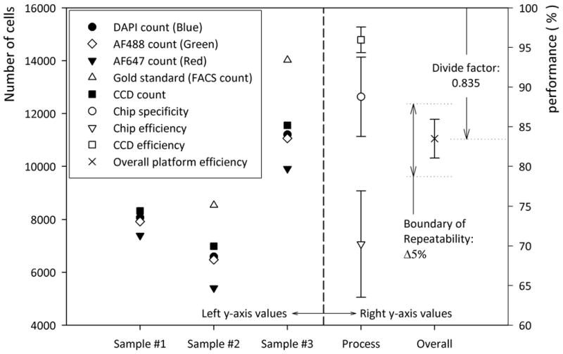
We demonstrate an integrated platform that merges a microfluidic chip with lensless imaging to target CD4(+) T-lymphocyte counts for HIV point-of-care testing at resource-limited settings. The chips were designed and fabricated simply with a laser cutter without using expensive cleanroom equipment. To capture CD4(+) T-lymphocytes from blood, anti-CD4 antibody was immobilized on only one side of the microfluidic chip. These captured cells were detected through an optically clear chip using a charge coupled device (CCD) sensor by lensless shadow imaging techniques. Gray scale image of the captured cells in a 24 mm x 4 mm x 50 microm microfluidic chip was obtained by the lensless imaging platform. The automatic cell counting software enumerated the captured cells in 3s. Captured cells were also imaged with a fluorescence microscope and manually counted to characterize functionality of the integrated platform. The integrated platform achieved 70.2+/-6.5% capture efficiency, 88.8+/-5.4% capture specificity for CD4(+) T-lymphocytes, 96+/-1.6% CCD efficiency, and 83.5+/-2.4% overall platform performance (n=9 devices) compared to the gold standard, i.e. flow cytometry count. The integrated system gives a CD4 count from blood within 10 min. The integrated platform points a promising direction for point-of-care testing (POCT) to rapidly capture, image and count subpopulations of cells from blood samples in an automated matter.


Figures

Figure 1
Figure 1
A schematic view of the CCD imaging platform: (a) CCD imaging platform to detect the captured cells. When light is incident on the captured cells, cells diffract and transmit light. Shadows of the captured CD4+ T-lymphocytes generated by diffraction can be imaged by the CCD in one second. Image is obtained with the lensless CCD imaging platform. (b) Picture of the microfluidic chip and CCD imaging platform. Field of view of the CCD sensor is 35 mm × 25 mm. The entire microfluidic device can be imaged without alignment by simply placing the microfluidic channel on the sensor. (c) Image taken with the lens-less CCD imaging platform and the shadow image of the cell in the microfluidic channel is shown. The image is obtained by diffraction. Scale bar, 100 μm.
Figure 1
Figure 1
A schematic view of the CCD imaging platform: (a) CCD imaging platform to detect the captured cells. When light is incident on the captured cells, cells diffract and transmit light. Shadows of the captured CD4+ T-lymphocytes generated by diffraction can be imaged by the CCD in one second. Image is obtained with the lensless CCD imaging platform. (b) Picture of the microfluidic chip and CCD imaging platform. Field of view of the CCD sensor is 35 mm × 25 mm. The entire microfluidic device can be imaged without alignment by simply placing the microfluidic channel on the sensor. (c) Image taken with the lens-less CCD imaging platform and the shadow image of the cell in the microfluidic channel is shown. The image is obtained by diffraction. Scale bar, 100 μm.
Figure 2
Figure 2
Cell capturing results using anti-CD4 antibody surface immobilization: (a) Optical and fluorescent images under fluorescent microscope to identify the captured cells. (i) Optical image of the channel (10×) (ii) DAPI stained cell image (UV excitation/blue emission) (iii) CD4+/AF488 stained cell image (blue excitation/green emission) (iv) CD3+/AF647 stained cell image (Orange excitation/red emission) were taken at the same position in microfluidic channel. All pictures have the same field of view. The flow direction is indicated, from inlet to outlet. Red arrows indicate artifacts detected by DAPI staining and white arrows indicate CD3-CD4+ cells, which were stained by anti-CD3 antibody-AF647 (Red). (b) Number of captured cells per unit area (mm2) in the microfluidic channel as a function of distance from the inlet. Scale bars are 5mm and 100 μm.
Figure 3
Figure 3
Graphical description for overall platform performance from table 1. To the left of graph the total cell count for three blood samples is shown. To the right of the graph the percentage values for chip efficiency, specificity, CCD efficiency, and overall platform efficiency are shown based on table 1. The boundary of repeatability indicates the conventional FACs error. The divide factor, “0.835”, indicates the correction factor based on overall platform efficiency.

References

    1. Cheng X, Irimia D, Dixon M, Sekine K, Demirci U, Zamir L, Tompkins RG, Rodriguez W, Toner M. Lab on a Chip. 2007a;7(2):170–178. - PMC - PubMed
    1. Cheng X, Irimia D, Dixon M, Ziperstein JC, Demirci U, Zamir L, Tompkins RG, Toner M, Rodriguez WR. Journal of acquired immune deficiency syndromes. 2007b;45(3):257–261. - PubMed
    1. Cheng X, Liu Ys, Irimia D, Demirci U, Yang L, Zamir L, Rodriguez WR, Toner M, Bashir R. Lab on a Chip. 2007c;7(6):746–755. - PMC - PubMed
    1. Chin CD, Linder V, Sia SK. Lab on a Chip. 2007;7(1):41–57. - PubMed
    1. Cui X, Lee LM, Heng X, Zhong W, Sternberg PW, Psaltis D, Yang C. Proceedings of the National Academy of Sciences. 2008;105(31):10670–10675. - PMC - PubMed

Publication types

MeSH terms