. 2009 Sep;132(Pt 9):2403-12.
doi: 10.1093/brain/awp125. Epub 2009 May 25.

Altered dopaminergic profile in the putamen and substantia nigra in restless leg syndrome

Affiliations

Altered dopaminergic profile in the putamen and substantia nigra in restless leg syndrome

James R Connor et al. Brain. 2009 Sep.

Abstract

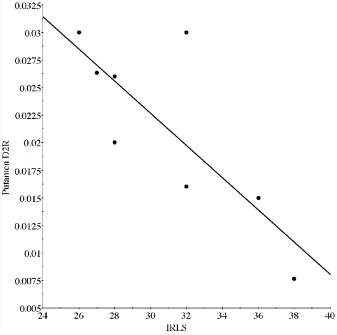
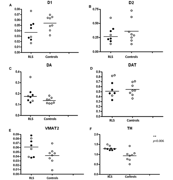
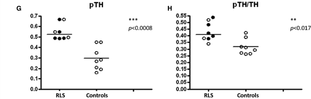
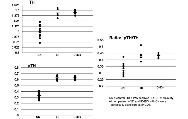
Restless leg syndrome (RLS) is a sensorimotor disorder. Clinical studies have implicated the dopaminergic system in RLS, while others have suggested that it is associated with insufficient levels of brain iron. To date, alterations in brain iron status have been demonstrated but, despite suggestions from the clinical literature, there have been no consistent findings documenting a dopaminergic abnormality in RLS brain tissue. In this study, the substantia nigra and putamen were obtained at autopsy from individuals with primary RLS and a neurologically normal control group. A quantitative profile of the dopaminergic system was obtained. Additional assays were performed on a catecholaminergic cell line and animal models of iron deficiency. RLS tissue, compared with controls, showed a significant decrease in D2R in the putamen that correlated with severity of the RLS. RLS also showed significant increases in tyrosine hydroxylase (TH) in the substantia nigra, compared with the controls, but not in the putamen. Both TH and phosphorylated (active) TH were significantly increased in both the substantia nigra and putamen. There were no significant differences in either the putamen or nigra for dopamine receptor 1, dopamine transporters or for VMAT. Significant increases in TH and phosphorylated TH were also seen in both the animal and cell models of iron insufficiency similar to that from the RLS autopsy data. For the first time, a clear indication of dopamine pathology in RLS is revealed in this autopsy study. The results suggest cellular regulation of dopamine production that closely matches the data from cellular and animal iron insufficiency models. The results are consistent with the hypothesis that a primary iron insufficiency produces a dopaminergic abnormality characterized as an overly activated dopaminergic system as part of the RLS pathology.


Figures

Figure 1
Figure 1
Dopaminergic profile in the putamen. The expression of D1 (A) D2 (B), dopamine; DA (C), DAT (D), VMAT2 (E), TH (F), pTH (G) and the pTH/TH ratio (H), in the putamen of control (right column in each graph) and RLS patients (left column in each graph). The data are plotted as individual values and the median is indicated by the bar in each graph. Those RLS patients who received dopaminergic agents as part of the treatment regimen are indicated by the closed circles and those who were not treated with dopaminergic agents are shown as open circles. Statistical significance was determined using the Mann–Whitney U-test.
Figure 2
Figure 2
Correlational analysis of putamenal D2 receptors and IRLS score. There was a strong inverse correlation (r = 0.80, P = 0.018) between the amount of D2 expression measured in the autopsy samples in the putamen and the patient's score on the IRLS scale of RLS severity. The higher the score on the rating scale indicating the more severe the symptoms.
Figure 3
Figure 3
Dopaminergic profile in the substantia nigra. The expression of D1 (A), D2 (B), dopamine; DA (C), DAT (D), VMAT (E), TH (F), pTH (G), and the pTH/TH ratio (H), in the substantia nigra of control (right column in each graph) and RLS patients (left column in each graph). The data are plotted as individual values and the median is indicated by the bar in each graph. Those RLS patients who received dopaminergic agents as part of the treatment regimen are indicated by the closed circles and those who were not treated with dopaminergic agents are shown as open circles. Statistical significance was determined using the Mann–Whitney U-test.
Figure 3
Figure 3
Dopaminergic profile in the substantia nigra. The expression of D1 (A), D2 (B), dopamine; DA (C), DAT (D), VMAT (E), TH (F), pTH (G), and the pTH/TH ratio (H), in the substantia nigra of control (right column in each graph) and RLS patients (left column in each graph). The data are plotted as individual values and the median is indicated by the bar in each graph. Those RLS patients who received dopaminergic agents as part of the treatment regimen are indicated by the closed circles and those who were not treated with dopaminergic agents are shown as open circles. Statistical significance was determined using the Mann–Whitney U-test.
Figure 4
Figure 4
TH and pTH. Immunoblot analysis for homogenates of rat ventral tegmentum (65-day-old rats). There is a significant increase in relative amounts of TH in rats that were exposed to a low-iron diet from gestational Day 5 through weaning at postnatal Day 21 and continued until 65 days (ID) compared with an age-matched control group (CN). Introducing an iron sufficient diet at the time of weaning did not result in a return to normal levels of TH by 65 days of age (ID–IDs). The levels of pTH and the ratio of pTH/TH followed the same patterns as described for TH. The differences for TH, pTH and the ratio from control are all significant (P < 0.001). The data are presented as X ± SE.
Figure 5
Figure 5
The effect of iron chelation on TH, pTH concentrations and pTH/TH ratios (pTH/TH) in PC12 cells. The concentrations of the iron chelator (DFO) are shown. Exposing cells to an iron chelator is associated with a trend of increasing amounts of TH, pTH and the pTH/TH ratio. The data are presented as X ± SE. Asterisk refers to significantly different compared with control.

References

    1. Allen R. Dopamine and iron in the pathophysiology of restless legs syndrome (RLS) Sleep Med. 2004;5:385–91. - PubMed
    1. Allen RP, Barker PB, Wehrl F, Song HK, Earley CJ. MRI measurement of brain iron in patients with restless legs syndrome. Neurology. 2001;56:263–5. - PubMed
    1. Allen RP, Connor JR, Hyland K, Earley CJ. Abnormally increased CSF 3-Ortho-methyldopa (3-OMD) in untreated restless legs syndrome (RLS) patients indicates more severe disease and possibly abnormally increased dopamine synthesis. Sleep Med. 2008;10:123–8. - PMC - PubMed
    1. Allen RP, Earley CJ. Defining the phenotype of the restless legs syndrome (RLS) using age-of-symptom-onset. Sleep Med. 2000;1:11–9. - PubMed
    1. Allen RP, Picchietti D, Hening WA, Trenkwalder C, Walters AS, Montplaisir J. Restless legs syndrome: diagnostic criteria, special considerations, and epidemiology. A report from the restless legs syndrome diagnosis and epidemiology workshop at the National Institutes of Health. Sleep Med. 2003;4:101–19. - PubMed

Publication types

MeSH terms

Substances