

Intermediate filaments exchange subunits along their length and elongate by end-to-end annealing
- PMID: 19468066
- PMCID: PMC2711597
- DOI: 10.1083/jcb.200809166
Intermediate filaments exchange subunits along their length and elongate by end-to-end annealing
Abstract
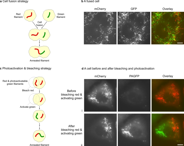
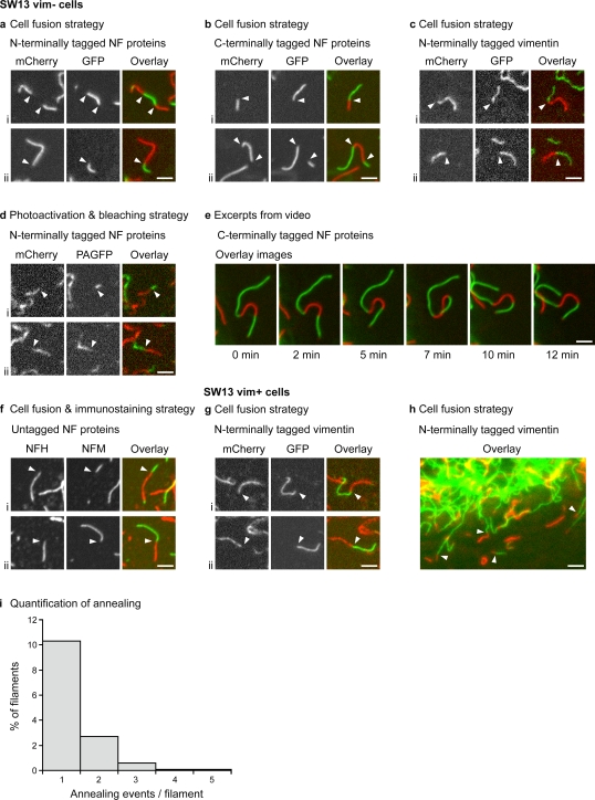
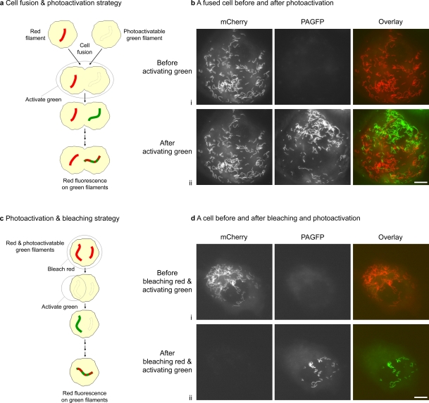
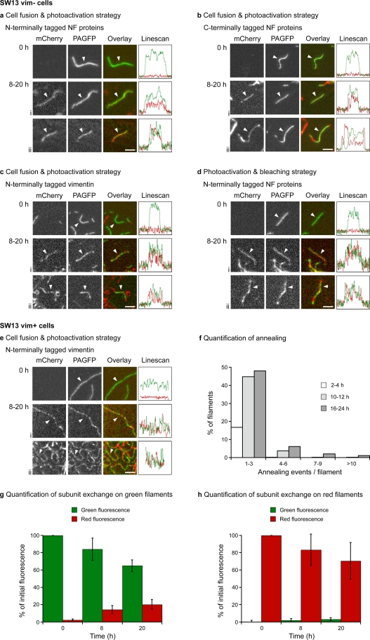
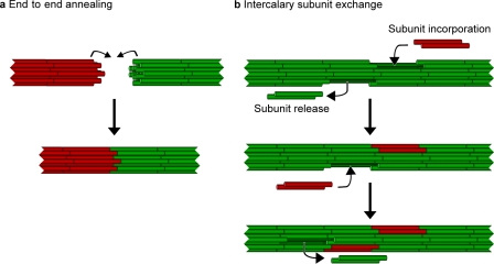
Actin filaments and microtubules lengthen and shorten by addition and loss of subunits at their ends, but it is not known whether this is also true for intermediate filaments. In fact, several studies suggest that in vivo, intermediate filaments may lengthen by end-to-end annealing and that addition and loss of subunits is not confined to the filament ends. To test these hypotheses, we investigated the assembly dynamics of neurofilament and vimentin intermediate filament proteins in cultured cells using cell fusion, photobleaching, and photoactivation strategies in combination with conventional and photoactivatable fluorescent fusion proteins. We show that neurofilaments and vimentin filaments lengthen by end-to-end annealing of assembled filaments. We also show that neurofilaments and vimentin filaments incorporate subunits along their length by intercalation into the filament wall with no preferential addition of subunits to the filament ends, a process which we term intercalary subunit exchange.
Figures
Similar articles
-
Severing and end-to-end annealing of neurofilaments in neurons.Proc Natl Acad Sci U S A. 2013 Jul 16;110(29):E2696-705. doi: 10.1073/pnas.1221835110. Epub 2013 Jul 2. Proc Natl Acad Sci U S A. 2013. PMID: 23821747 Free PMC article.
-
Molecular architecture of the neurofilament. II. Reassembly process of neurofilament L protein in vitro.J Mol Biol. 1990 Feb 20;211(4):871-82. doi: 10.1016/0022-2836(90)90080-6. J Mol Biol. 1990. PMID: 2313699
-
Characterization of distinct early assembly units of different intermediate filament proteins.J Mol Biol. 1999 Mar 12;286(5):1403-20. doi: 10.1006/jmbi.1999.2528. J Mol Biol. 1999. PMID: 10064706
-
Role of phosphorylation on the structural dynamics and function of types III and IV intermediate filaments.Exp Cell Res. 2007 Jun 10;313(10):2098-109. doi: 10.1016/j.yexcr.2007.04.010. Epub 2007 Apr 12. Exp Cell Res. 2007. PMID: 17498690 Free PMC article. Review.
-
The third wave: Intermediate filaments in the maturing nervous system.Mol Cell Neurosci. 2017 Oct;84:68-76. doi: 10.1016/j.mcn.2017.05.010. Epub 2017 May 26. Mol Cell Neurosci. 2017. PMID: 28554564 Review.
Cited by
-
The role of neurofilament aggregation in neurodegeneration: lessons from rare inherited neurological disorders.Mol Neurodegener. 2019 May 16;14(1):19. doi: 10.1186/s13024-019-0318-4. Mol Neurodegener. 2019. PMID: 31097008 Free PMC article. Review.
-
Molecular insights into cardiomyopathies associated with desmin (DES) mutations.Biophys Rev. 2018 Aug;10(4):983-1006. doi: 10.1007/s12551-018-0429-0. Epub 2018 Jun 20. Biophys Rev. 2018. PMID: 29926427 Free PMC article. Review.
-
Endoplasmic spreading requires coalescence of vimentin intermediate filaments at force-bearing adhesions.Mol Biol Cell. 2013 Jan;24(1):21-30. doi: 10.1091/mbc.E12-05-0377. Epub 2012 Oct 31. Mol Biol Cell. 2013. PMID: 23115305 Free PMC article.
-
Axonal neurofilaments exhibit frequent and complex folding behaviors.Cytoskeleton (Hoboken). 2018 Jun;75(6):258-280. doi: 10.1002/cm.21448. Cytoskeleton (Hoboken). 2018. PMID: 29683261 Free PMC article.
-
Direct observation of subunit exchange along mature vimentin intermediate filaments.Biophys J. 2014 Dec 16;107(12):2923-2931. doi: 10.1016/j.bpj.2014.09.050. Biophys J. 2014. PMID: 25517157 Free PMC article.
References
-
- Alberts B., Johnson A., Lewis J., Raff M., Roberts K., Walter P. 2002. Molecular Biology of the Cell. Fourth edition Garland Science, New York: 1548 pp
-
- Andrianantoandro E., Blanchoin L., Sept D., McCammon J.A., Pollard T.D. 2001. Kinetic mechanism of end-to-end annealing of actin filaments.J. Mol. Biol. 312:721–730 - PubMed
-
- Burton P.R., Wentz M.A. 1992. Neurofilaments are prominent in bullfrog olfactory axons but are rarely seen in those of the tiger salamander, Ambystoma tigrinum.J. Comp. Neurol. 317:396–406 - PubMed
Publication types
MeSH terms
Substances
Grants and funding
LinkOut - more resources
Full Text Sources
Other Literature Sources
Research Materials
