

Induction of alternative lengthening of telomeres-associated PML bodies by p53/p21 requires HP1 proteins
- PMID: 19468068
- PMCID: PMC2711592
- DOI: 10.1083/jcb.200810084
Induction of alternative lengthening of telomeres-associated PML bodies by p53/p21 requires HP1 proteins
Abstract
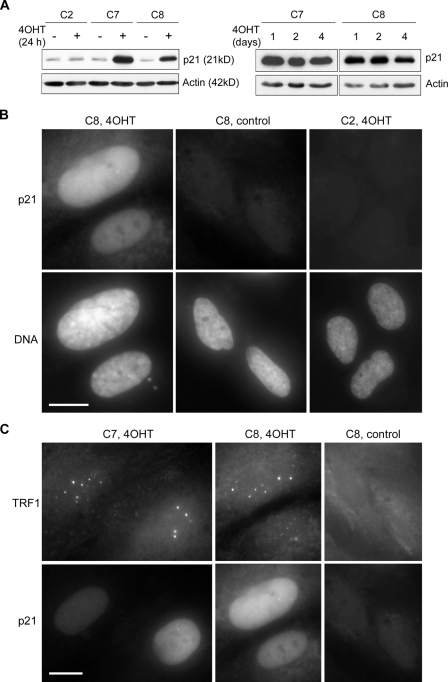
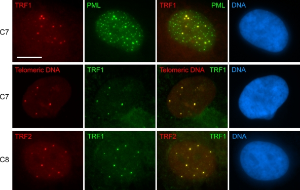
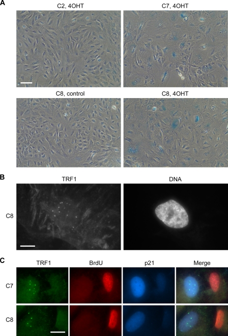
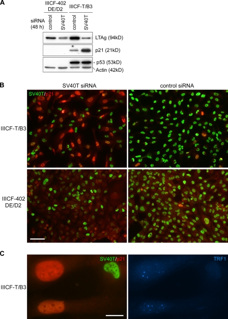
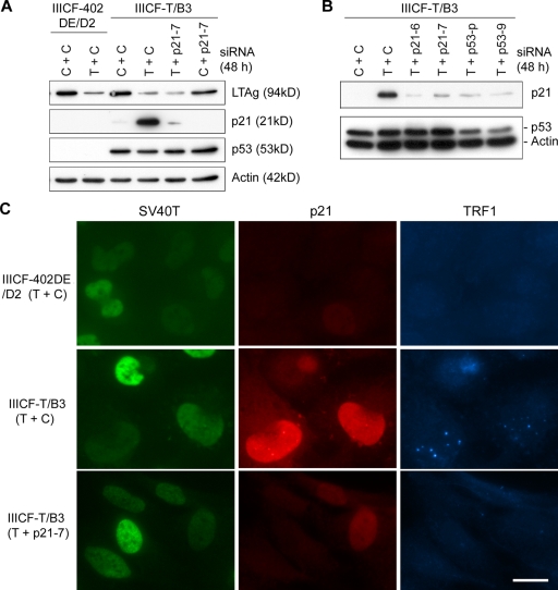
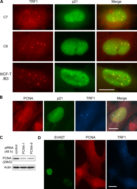
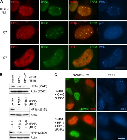
Alternative lengthening of telomeres (ALT) is a recombination-mediated process that maintains telomeres in telomerase-negative cancer cells. In asynchronously dividing ALT-positive cell populations, a small fraction of the cells have ALT-associated promyelocytic leukemia nuclear bodies (APBs), which contain (TTAGGG)n DNA and telomere-binding proteins. We found that restoring p53 function in ALT cells caused p21 up-regulation, growth arrest/senescence, and a large increase in cells containing APBs. Knockdown of p21 significantly reduced p53-mediated induction of APBs. Moreover, we found that heterochromatin protein 1 (HP1) is present in APBs, and knockdown of HP1alpha and/or HP1gamma prevented p53-mediated APB induction, which suggests that HP1-mediated chromatin compaction is required for APB formation. Therefore, although the presence of APBs in a cell line or tumor is an excellent qualitative marker for ALT, the association of APBs with growth arrest/senescence and with "closed" telomeric chromatin, which is likely to repress recombination, suggests there is no simple correlation between ALT activity level and the number of APBs or APB-positive cells.
Figures
Similar articles
-
HP1-mediated formation of alternative lengthening of telomeres-associated PML bodies requires HIRA but not ASF1a.PLoS One. 2011 Feb 15;6(2):e17036. doi: 10.1371/journal.pone.0017036. PLoS One. 2011. PMID: 21347226 Free PMC article.
-
Identification of candidate alternative lengthening of telomeres genes by methionine restriction and RNA interference.Oncogene. 2007 Jul 12;26(32):4635-47. doi: 10.1038/sj.onc.1210260. Epub 2007 Feb 5. Oncogene. 2007. PMID: 17297460
-
p53 and p21(Waf1) are recruited to distinct PML-containing nuclear foci in irradiated and Nutlin-3a-treated U2OS cells.J Cell Biochem. 2010 Dec 1;111(5):1280-90. doi: 10.1002/jcb.22852. J Cell Biochem. 2010. PMID: 20803550 Free PMC article.
-
Alternative lengthening of telomeres in mammalian cells.Oncogene. 2002 Jan 21;21(4):598-610. doi: 10.1038/sj.onc.1205058. Oncogene. 2002. PMID: 11850785 Review.
-
PML body meets telomere: the beginning of an ALTernate ending?Nucleus. 2012 May-Jun;3(3):263-75. doi: 10.4161/nucl.20326. Epub 2012 May 1. Nucleus. 2012. PMID: 22572954 Free PMC article. Review.
Cited by
-
Telomeric armor: the layers of end protection.J Cell Sci. 2009 Nov 15;122(Pt 22):4013-25. doi: 10.1242/jcs.050567. J Cell Sci. 2009. PMID: 19910493 Free PMC article.
-
Recruitment of TRIM33 to cell-context specific PML nuclear bodies regulates nodal signaling in mESCs.EMBO J. 2023 Feb 1;42(3):e112058. doi: 10.15252/embj.2022112058. Epub 2022 Dec 16. EMBO J. 2023. PMID: 36524443 Free PMC article.
-
Nuclear bodies: random aggregates of sticky proteins or crucibles of macromolecular assembly?Dev Cell. 2009 Nov;17(5):639-47. doi: 10.1016/j.devcel.2009.10.017. Dev Cell. 2009. PMID: 19922869 Free PMC article. Review.
-
Alternative lengthening of telomeres: models, mechanisms and implications.Nat Rev Genet. 2010 May;11(5):319-30. doi: 10.1038/nrg2763. Epub 2010 Mar 30. Nat Rev Genet. 2010. PMID: 20351727 Review.
-
Molecular mechanisms of activity and derepression of alternative lengthening of telomeres.Nat Struct Mol Biol. 2015 Nov;22(11):875-80. doi: 10.1038/nsmb.3106. Epub 2015 Nov 4. Nat Struct Mol Biol. 2015. PMID: 26581522 Review.
References
-
- Blasco M.A. 2007. The epigenetic regulation of mammalian telomeres.Nat. Rev. Genet. 8:299–309 - PubMed
-
- Brachner A., Sasgary S., Pirker C., Rodgarkia C., Mikula M., Mikulits W., Bergmeister H., Setinek U., Wieser M., Chin S.F., et al. 2006. Telomerase- and alternative telomere lengthening-independent telomere stabilization in a metastasis-derived human non-small cell lung cancer cell line: effect of ectopic hTERT.Cancer Res. 66:3584–3592 - PubMed
-
- Brown J.P., Wei W., Sedivy J.M. 1997. Bypass of senescence after disruption of p21CIP1/WAF1 gene in normal diploid human fibroblasts.Science. 277:831–834 - PubMed
-
- Bryan T.M., Englezou A., Dalla-Pozza L., Dunham M.A., Reddel R.R. 1997. Evidence for an alternative mechanism for maintaining telomere length in human tumors and tumor-derived cell lines.Nat. Med. 3:1271–1274 - PubMed
Publication types
MeSH terms
Substances
LinkOut - more resources
Full Text Sources
Other Literature Sources
Research Materials
Miscellaneous
