. 2009 Jun 1;185(5):889-902.
doi: 10.1083/jcb.200807066. Epub 2009 May 25.

A trans-membrane segment inside the ribosome exit tunnel triggers RAMP4 recruitment to the Sec61p translocase

Affiliations

A trans-membrane segment inside the ribosome exit tunnel triggers RAMP4 recruitment to the Sec61p translocase

Martin R Pool. J Cell Biol. .

Abstract

Membrane protein integration occurs predominantly at the endoplasmic reticulum and is mediated by the translocon, which is formed by the Sec61p complex. The translocon binds to the ribosome at the polypeptide exit site such that integration occurs in a cotranslational manner. Ribosomal protein Rpl17 is positioned such that it contacts both the ribosome exit tunnel and the surface of the ribosome near the exit site, where it is intimately associated with the translocon. The presence of a trans-membrane (TM) segment inside the ribosomal exit tunnel leads to the recruitment of RAMP4 to the translocon at a site adjacent to Rpl17. This suggests a signaling function for Rpl17 such that it can recognize a TM segment inside the ribosome and triggers rearrangements of the translocon, priming it for subsequent TM segment integration.


Figures

Figure 1.
Figure 1.
Membrane components protect Rpl17 from proteolytic digestion. (A) Free ribosomes and RMs were treated with increasing concentrations of V8 protease and analyzed by SDS-PAGE and immunoblotting with Rpl17N-trm, Rpl18, and Rpl23a antibodies. The position of a stable Rpl17 degradation product, which lacks the C terminus (Rpl17ΔC), is indicated. The antibody weakly cross reacts with another ribosomal protein (*). (B) RMs were solubilized with 2% digitonin, the membranes were subjected to limited digestion with V8 protease, and analyzed as described in A. A mock-treated sample (−digitonin) was treated in an identical manner but with no digitonin present.
Figure 2.
Figure 2.
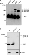
Cross-linking reveals that Rpl17 is adjacent to membrane components. (A) RMs were treated with either DMSO or the cross-linkers DFDNB (25 µM), DSG (200 µM), and MBS (200 µM). Reactions were analyzed by SDS-PAGE and immunoblotting with Rpl17C-trm antibodies. Major cross-links to adducts of the approximate indicated sizes are labeled. An additional, weak 7-kD cross-link adduct (o) was also reproducibly observed with MBS. (B) RMs were treated with 200 µM MBS to induce cross-linking. The microsomes were treated with 1 M LiCl to extract ribosomal proteins from the rRNA and separated from the ribosomal remnants by floatation through a Nycodenz gradient. The floated and nonfloated material were recovered by TCA precipitation and analyzed by SDS-PAGE and immunoblotting with Rpl17C-trm antibodies. To control for complete floatation of the microsomes, the blot was also probed with antibodies against SRβ, an integral ER membrane protein.
Figure 3.
Figure 3.
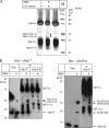
Rpl17 can be cross-linked to Sec61β. (A) RMs (900 eq) were cross-linked where indicated with 200 µM MBS. After solubilization with digitonin, ribosomes were reisolated, ribosomal proteins were extracted with LiCl, and the extract was immunoprecipitated with anti-Rpl17C-trm antibodies. Bound protein was eluted with SDS and analyzed by SDS-PAGE and staining with Coomassie brilliant blue. A mock immunoprecipitation performed in the absence of a microsomal extract was also performed to identify bands arising from the antiserum. The prominent band at 21 kD (*) was excised along with the 30-kD band that was exclusive to the LiCl extract from MBS-treated RM (♦) and analyzed by mass spectrometry after in-gel tryptic digestion. M, molecular weight marker. (B) Denaturing immunoprecipitation (IP) of cross-linking reactions (100 eq of RM) was performed as above either using anti-Rpl17C-trm or anti-Sec61β antiserum. The samples were analyzed by SDS-PAGE and immunoblotting using Rpl17C-trm antiserum (left) or Sec61β antiserum (right). Position of IgG heavy chain (hc) and light chains (lc) are indicated. White lines indicate that intervening lanes have been spliced out.
Figure 4.
Figure 4.
Sec61β and Rpl17 remain in proximity after membrane solubilization. RMs were resuspended in solubilization buffer and where indicated solubilized with 2% digitonin. Cross-linking was induced with either MBS or DSG. The reactions were analyzed by SDS-PAGE and immunoblotting with Sec61β antisera. Two major cross-link species, indicated by asterisks, were only present in intact membranes and were lost after detergent treatment.
Figure 5.
Figure 5.
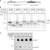
Generation and purification of pPV integration intermediates. (A) Schematic representation of pPV fusion protein, consisting of the N terminus of pPL, including the signal sequence (SS), fused to the TM domain (TMD) segment and C-terminal cytosolic domain of VSV-G. (B) mRNAs encoding pPL and pPV fusion proteins of defined lengths, as indicated, and lacking a stop codon together with full-length pPV with an intact stop codon (pPV116STOP) were translated in rabbit reticulocyte lysate in the presence of EKRM and purified SRP. Where indicated, the reactions were treated with puromycin to release the nascent chain from the ribosome. The samples were precipitated and analyzed by SDS-PAGE and phosphorimaging. The unprocessed pPL/pPV (*) and signal sequence–processed PL/PV (•) forms are indicated. (C) Where indicated, insertion reactions were programmed with pPV mRNAs of the indicated length lacking a stop codon. The resulting translocation intermediates were stabilized with cycloheximide and adjusted to 500 mM KOAc. The membranes were reisolated by floatation through an HS Nycodenz gradient, and the floated (F) and unfloated (U) fractions were collected. Fractions were analyzed by SDS-PAGE and immunoblotting with Rpl17N-trm and Sec61β antisera.
Figure 6.
Figure 6.
Cross-linking of Rpl17 in pPV integration intermediates. (A) Insertion reactions were performed using pPV mRNAs of defined nascent chain (nc) lengths. As a control, a mock insertion reaction lacking exogenous mRNA was also performed (−). The resulting integration intermediates were purified by floatation through an HS gradient (as described in Fig. 5 C) and treated with 200 µM MBS before analysis by SDS-PAGE and immunoblotting for Rpl17C-trm. (B) Translocation intermediates were generated as in A using pPL86mer or 110mer, and these were purified and treated with MBS. (C) Translocation intermediates were generated and treated with MBS as in A using a mutant of pPV in which seven of the hydrophobic residues within the TM segment were mutated to polar residues as indicated (^). (D) RM were treated either with DMSO (−) or 200 µM MBS and analyzed by SDS-PAGE and immunoblotting for Rpl17C-trm. A long exposure of the immunoblot reveals, in addition to the two Sec61β cross-link products, two weaker cross-link products of ∼6 and ∼7 kD. White line indicates that intervening lanes have been spliced out.
Figure 7.
Figure 7.
The presence of a TM segment leads to differential cross-linking between Rpl17 and RAMP4. (A) RMs were either mock treated or treated with 25 mM EDTA before cross-linking with 200 µM MBS. The reactions were separated on the same SDS-PAGE gel and immunoblotted for Rpl17C-trm, RAMP4, and Sec61β. Positions of the Rpl17-Sec61β cross-links are indicated (*) together with the 7-kD Rpl17 cross-link, which comigrates with a 20-kD RAMP4 cross-link (>). (B) Denaturing immunoprecipitation (IP) of cross-linking reactions (100 eq of RM) was performed using either anti-Rpl17C-trm or anti-RAMP4 antiserum. The samples were analyzed by SDS-PAGE and immunoblotting using either anti-RAMP4 or anti-Rpl17C-trm antibodies. Total and immunoprecipitation fractions were lanes from the same gel with exposure times of 30 s and 3 min, respectively. The position of 27-kD Rpl17-RAMP4 cross-link product is indicated (o). (C) pPV translocation intermediates of defined nascent chain (nc) lengths (residues) were generated, purified, and treated with MBS as described in Fig. 6 A.The reactions were analyzed by SDS-PAGE and immunoblotting with RAMP4 antisera. The 27-kD Rpl17-RAMP4 cross-link product is again indicated and shows a strong dependence on the length of the pPV nascent chain. Values on blots are shown in kilodaltons. White lines indicate that intervening lanes have been spliced out.
Figure 8.
Figure 8.
RAMP4 is recruited to the ribosome–translocon complex. (A) RMs were solubilized with digitonin and centrifuged through a sucrose cushion to yield a ribosomal pellet that contains RAMPs. Equal amounts of the total and RAMP fraction were analyzed by SDS-PAGE and immunoblotting with Rpl17, Sec61β, and RAMP4 antisera. (B) Cross-linking of microsomes with MBS before and after digitonin treatment was performed as described in Fig. 4. Reactions were analyzed by immunoblotting with Rpl17C-trm antiserum. Position of the 27-kD Rpl17xRAMP4 cross-link species is indicated (o). Values on blot are shown in kilodaltons. (C) pPV translocation intermediates with chain lengths of 87 and 94 were generated and purified as described in Fig. 6 A, and they were solubilized with digitonin and the RAMP fraction prepared as in A. Fractions were analyzed, alongside 5% of the input PKRM used to generate the translocation intermediates, by immunoblotting with Rpl17, Sec61β, and RAMP4 antisera.

Comment in

References

    1. Bacher G., Pool M., Dobberstein B. 1999. The ribosome regulates the GTPase of the β-subunit of the signal recognition particle receptor.J. Cell Biol. 146:723–730 - PMC - PubMed
    1. Ban N., Nissen P., Hansen J., Moore P.B., Steitz T.A. 2000. The complete atomic structure of the large ribosomal subunit at 2.4 Å.Science. 289:905–920 - PubMed
    1. Beckmann R., Spahn C.M.T., Eswar N., Helmers J., Penczek P.A., Sall A., Frank J., Blobel G. 2001. Architecture of the protein-conducting channel associated with the translating 80S ribosome.Cell. 107:361–372 - PubMed
    1. Besemer J., Harant H., Wang S., Oberhauser B., Marquardt K., Foster C.A., Schreiner E.P., de Vries J.E., Dascher-Nadel C., Lindley I.J. 2005. Selective inhibition of cotranslational translocation of vascular cell adhesion molecule 1.Nature. 436:290–293 - PubMed
    1. Bornemann T., Jockel J., Rodnina M.V., Wintermeyer W. 2008. Signal sequence-independent membrane targeting of ribosomes containing short nascent peptides within the exit tunnel.Nat. Struct. Mol. Biol. 15:494–499 - PubMed