. 2009 May;5(5):e1000435.
doi: 10.1371/journal.ppat.1000435. Epub 2009 May 15.

Intrinsic thermal sensing controls proteolysis of Yersinia virulence regulator RovA

Affiliations

Intrinsic thermal sensing controls proteolysis of Yersinia virulence regulator RovA

Katharina Herbst et al. PLoS Pathog. 2009 May.

Abstract

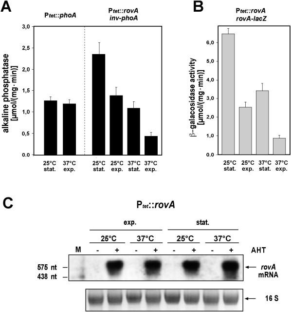
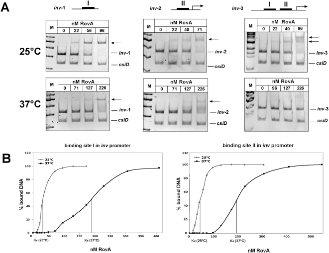
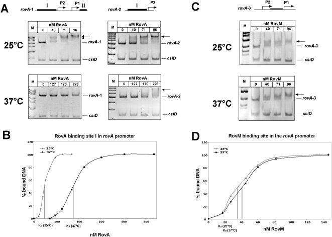
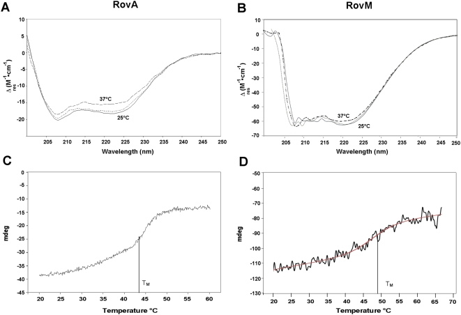
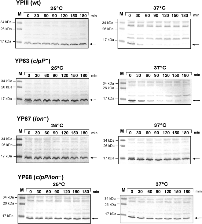
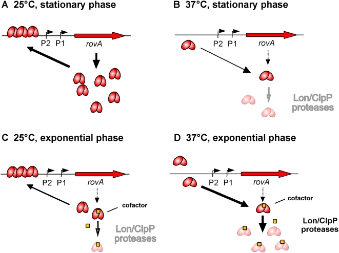
Pathogens, which alternate between environmental reservoirs and a mammalian host, frequently use thermal sensing devices to adjust virulence gene expression. Here, we identify the Yersinia virulence regulator RovA as a protein thermometer. Thermal shifts encountered upon host entry lead to a reversible conformational change of the autoactivator, which reduces its DNA-binding functions and renders it more susceptible for proteolysis. Cooperative binding of RovA to its target promoters is significantly reduced at 37 degrees C, indicating that temperature control of rovA transcription is primarily based on the autoregulatory loop. Thermally induced reduction of DNA-binding is accompanied by an enhanced degradation of RovA, primarily by the Lon protease. This process is also subject to growth phase control. Studies with modified/chimeric RovA proteins indicate that amino acid residues in the vicinity of the central DNA-binding domain are important for proteolytic susceptibility. Our results establish RovA as an intrinsic temperature-sensing protein in which thermally induced conformational changes interfere with DNA-binding capacity, and secondarily render RovA susceptible to proteolytic degradation.


Conflict of interest statement

The authors have declared that no competing interests exist.

Figures

Figure 1
Figure 1. Temperature and growth phase-dependent rovA expression occurs on the post-transcriptional level.
(A, left panel) Analysis of the expression of the phoA gene under the control of the tetracycline promoter (Ptet). Quantification of the expression of the inv-phoA fusion (A, right panel), and the rovA-lacZ fusion (B) in E. coli strain DH5αZ1 pHT123, in which rovA is being expressed under the control of the tetracycline promoter (Ptet). Strain DH5αZ1, encoding the tet repressor gene tetR, was grown overnight or to exponential phase at 25°C or 37°C in the presence of the inducer AHT (0,1 µg ml−1). β-galactosidase and alkaline phosphatase activity is the mean of at least three independent determinations done in triplicate ±SD. (C) DH5αZ1 pHT123 was grown in the presence (+) or absence (−) of the inducer AHT 0,1 µg ml−1 under indicated growth conditions. Total RNA was prepared, separated on a 0.7% agarose gel, transferred onto a Nylon membrane and probed with a digoxigenin (DIG)-labelled PCR fragment encoding the rovA gene. 16 rRNA was used as a loading control. A DIG-labelled RNA marker is loaded in the left lane (M), and arrows indicate the rovA mRNA transcript.
Figure 2
Figure 2. Interaction of RovA with the inv regulatory region at 25°C or 37°C.
(A) The double-stranded promoter fragments of the inv regulatory region harbouring all RovA binding sites, or only the binding site(s) in region I or II (black boxes) were incubated without or with rising amounts of purified RovA protein at 25°C or 37°C. The DNA-RovA complexes were separated on a 4% polyacrylamide gel. A non-specific probe containing an unrelated sequence (csiD promoter of E. coli) was included as negative control. Different DNA fragments used for the band shift assays are illustrated on top. Bold lines indicate previously identified RovA binding sites. Band shift analyses of the chosen fragments are shown below. (B) A constant concentration of DNA fragments, harbouring site I or II of the inv promoter region, were incubated with different increasing concentrations of RovA and subjected to band shift assays. The DNA bands were quantified and the Kd was defined as the protein concentration required for half-maximal binding and was calculated for each protein-DNA concentration and presented as average of at least four independent experiments.
Figure 3
Figure 3. Interaction of RovA or RovM with the rovA regulatory region at 25°C or 37°C.
Double-stranded promoter fragments of the rovA regulatory region harbouring RovA binding sites (black boxes) or the RovM binding site (grey box) were incubated without or with rising amounts of purified RovA protein (A) or RovM (C) at 25°C or 37°C. The DNA-protein complexes were separated on a 4% polyacrylamide gel. A non-specific probe containing an unrelated sequence (csiD promoter of E. coli) was included as negative control. Different DNA fragments used for the band shift assays are illustrated on top. Bold lines indicate previously identified RovA or RovM binding sites. Band shift analyses of the chosen fragments are shown below. (B, D) A constant concentration of DNA fragments, harbouring RovA binding site I or the RovM binding site of the rovA promoter region, were incubated with different increasing concentrations of RovA (B), or RovM (D) and subjected to band shift assays. The DNA bands were quantified and the Kd was defined as the protein concentration required for half-maximal binding and was calculated for each protein-DNA concentration and presented as average of at least four independent experiments.
Figure 4
Figure 4. Conformational analysis of RovA and RovM using CD spectroscopy.
CS spectra, Δε (M−1 cm−1) versus wavelength of RovA (0.16 mg/ml) (A) or RovM (0.4 mg/ml) (B) as function of temperature (25°C, solid line; 37°C broken line; 37°C and cooling to 25°C dotted line). Thermal stability of the RovA (C) and the RovM (D) protein. The temperature of the purified protein solutions were increased from 20°C to 60°C with a temperature slope of 2°C/min. The denaturation curves were recorded at fixed wavelength of λ = 222 nm. The melting points (TM) were calculated using the Jascow spectra analysis software.
Figure 5
Figure 5. Analysis of the intracellular RovA and RovM levels when expressed under the control of the tetracycline promoter (Ptet).
Y. pseudotuberculosis strain YP3 pHT123 (A), E. coli strain DH5αZ1 pHT123 (B) and E. coli strain DH5αZ1 pKH31 (C) were grown overnight or to exponential phase at 25°C or 37°C in the presence of the inducer AHT (0,1 µg ml−1). Whole cell extracts from the cultures were prepared, and analyzed by Western blotting with a polyclonal antibody directed against RovA or RovM .
Figure 6
Figure 6. Stability of RovA during exponential and stationary phase at 25°C and 37°C.
Cultures of Y. pseudotuberculosis strain YPIII (wt) (A–B) and E. coli strain DH5αZ1 pHT123 (C–F) were grown overnight or to exponential phase (OD600 = 0.3–0.4) at 25°C before chloramphenicol (200 µg ml−1) was added. The cultures were divided and incubated at 25°C or 37°C for additional 90 min. Aliquots of the cultures were removed at the indicated times thereafter, whole cell extracts from identical numbers of bacteria were prepared and analyzed by Western blotting with a polyclonal antibody directed against RovA. A whole cell extract from the rovA mutant strain YP3 grown overnight at 25°C was used as control. (E, F) Protein bands of DH5αZ1 pHT123 were quantified using the BioRad analysis software ‘Quantity One’ 4.6.2 and set into relation to sampling at time point zero (◆ 25°C, stationary phase; ▲ 37°C, stationary phase; ■ 25°C, exponential phase; ● 37°C, exponential phase).
Figure 7
Figure 7. RovA expression in Y. pseudotuberculosis wild-type and protease mutants at 25°C and 37°C.
Expression of a rovA-lacZ fusion on pAKH47 was analyzed in Y. pseudotuberculosis YPIII (wt) and in the clpP, lon and clpP/lon mutant strains (YP63, YP67 and YP68), when rovA was expressed from its own promoter (ProvA::rovA) (A), and in the presence of the rovA expression plasmid pHT123 (Ptet::rovA) (B). Cultures were grown at 25°C and 37°C in LB medium overnight and β-galactosidase activity was determined. The data represent the average ±SD from at least three different experiments each done in duplicate (upper panel). Furthermore, whole cell extracts of equal amounts of the bacteria were prepared, separated by SDS-PAGE, and visualized by immunoblotting using a polyclonal antibody directed against RovA.
Figure 8
Figure 8. Stability of RovA during exponential phase at 25°C and 37°C in Y. pseudotuberculosis YPIII and the clpP, lon, and clp/lon deletion mutants YP63, YP67 and YP68.
Cultures of Y. pseudotuberculosis strain YPIII (wt) and the clpP, lon, and clp/lon deletion mutants harbouring the Ptet::rovA expression plasmid pHT123 were grown to exponential phase (OD600 = 0.3–0.5) at 25°C before chloramphenicol (200 µg ml−1) was added. The cultures were divided and incubated at 25°C or 37°C for additional 180 min. Aliquots of the cultures were removed at the indicated times thereafter, whole cell extracts from identical number of bacteria were prepared and analyzed by Western blotting with a polyclonal antibody directed against RovA. The arrow indicates the RovA protein.
Figure 9
Figure 9. Stability of different RovA-LacZ fusion proteins during exponential phase at 37°C.
Cultures of Y. pseudotuberculosis strain YPIII (wt) harbouring the rovA-lacZ expression plasmid pGN17 (A), pKH16 (B), and pKH21-23 (C–E) and the lon deletion mutant YP67 harbouring the rovA-lacZ expression plasmid pGN17 (A) were grown to exponential phase (OD600 = 0.3–0.4) at 25°C before chloramphenicol (200 µg ml−1) was added. The cultures were shifted to 37°C for additional 90 min. Aliquots of the cultures were removed at the indicated times, whole cell extracts from identical numbers of bacteria were prepared and analyzed by Western blotting with a polyclonal antibody directed against β-galactosidase (A) or RovA (B–E). Structure of the RovA protein is given in the upper panel on the left. The predicted α-helical domains implicated in dimerization are given in dark grey, the two α-helices of the HTH motif are indicated by light grey boxes, and the two β-sheets of the winged HTH DNA-binding motif are given in light grey arrows. The numbers above the arrows indicate the last amino acid fused to β-galactosidase.
Figure 10
Figure 10. In vitro degradation of α-casein and RovA by Lon.
α-casein and purified RovA were incubated at 25°C and 37°C in the presence of ATP and an ATP-regeneration system without (A, C) or with (B, D) purified Lon. Whole cell extracts of BL21 (lon ) were prepared and added to purified Lon and RovA in the absence (E) and presence of ATP and the ATP regeneration system (F). The degradation reactions were performed as described in Material and Methods. An aliquot was removed at indicated times, separated on 15% SDS-gels, and visualised after staining with Coomassie brilliant blue (A–D) or by Western blotting with an anti-RovA antibody (E–F). * indicates protein bands that interacted non-specifically with the RovA antibody used for loading controls.
Figure 11
Figure 11. Model of temperature-mediated regulation of rovA expression.
(A) At moderate temperatures during stationary phase RovA is active and binds cooperatively to its operator sequences upstream of the rovA gene. DNA interaction of RovA stimulates transcription by direct activation of the RNA polymerase and antirepression of H-NS mediated silencing. Active conformation and/or DNA-binding block degradation by the Lon and ClpP proteases. (B) Elevated temperature (37°C) induces a conformational change to the inactive, non-DNA-binding form of RovA. This shift in the equilibrium favours the recognition and/or access by the proteases, which leads to an increased degradation of RovA by Lon and to a smaller extent also by ClpP. (C) During exponential phase, a portion of RovA (e.g. non-DNA bound protein) is destablized, e.g. by binding of a cofactor, which increases susceptibility and proteolysis by the proteases at moderate temperatures. (D) At 37°C, partial defolding and inactivation of the RovA protein further improves cofactor binding and/or allow a better access of the proteases, leading to rapid degradation of the regulatory protein.

Similar articles

Cited by

References

    1. Konkel ME, Tilly K. Temperature-regulated expression of bacterial virulence genes. Microbes Infect. 2000;2:157–166. - PubMed
    1. Schumann W. Thermosensors in eubacteria: role and evolution. J Biosci. 2007;32:549–557. - PubMed
    1. Straley SC, Perry RD. Environmental modulation of gene expression and pathogenesis in Yersinia. Trends Microbiol. 1995;3:310–317. - PubMed
    1. Stenseth NC, Atshabar BB, Begon M, Belmain SR, Bertherat E, et al. Plague: past, present, and future. PLoS Med. 2008;5:e3. doi:10.1371/journal.pmed.0050003. - PMC - PubMed
    1. Bottone EJ. Yersinia enterocolitica: the charisma continues. Clin Microbiol Rev. 1997;10:257–276. - PMC - PubMed

Publication types

MeSH terms