Perineal sound recording for diagnosis of bladder outlet obstruction
- PMID: 19468437
- PMCID: PMC2684325
- DOI: 10.4103/0970-1591.45545
Perineal sound recording for diagnosis of bladder outlet obstruction
Abstract
Objectives: Elderly men are prone to developing lower urinary tract symptoms (LUTS) possibly caused by bladder outlet obstruction (BOO). The most frequently used method to diagnose this condition is an invasive and time-consuming pressure-flow study. We are developing a novel non-invasive method to diagnose BOO in men with LUTS based on perineal sound recording.
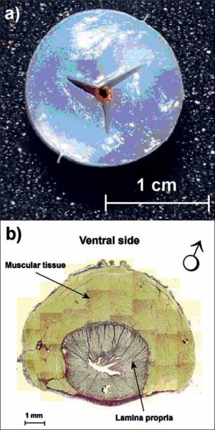
Methods: A biophysical model urethra was made from polyvinyl alcohol (PVA) cryogel with viscoelastic properties comparable to those of the male pig urethra. To this model different degrees of obstruction were applied and sound was recorded at different positions downstream of the obstruction. In a study in 16 healthy male volunteers the variability and repeatability of perineal sound recording was tested.
Results: In the model three parameters, derived from the frequency spectrum of the recorded sound (i.e., weighted average frequency, standard deviation and skewness) are uniquely related to the degree of obstruction (linear regression, P<0.001). The variability of perineal sound recording in healthy male volunteers was found to be smaller within volunteers than between volunteers (Kruskal-Wallis, P<0.001) and the repeatability was comparable to that of the maximum flow rate.
Conclusions: We conclude that perineal sound recordings are significantly different between volunteers. In combination with the unique relations found in the model-experiments these results increase the probability that perineal sound recording can be used as a simple and cheap non-invasive method to diagnose BOO. Clinical testing of this method is therefore strongly indicated.
Keywords: Bladder outlet obstruction; non-invasive urodynamics; perineal sound recording.
Conflict of interest statement
Figures


References
-
- Griffiths DJ, Höfner K, van Mastrigt R, Rollema HJ, Spångberg A, Gleason DM. Standardization of terminology of lower urinary tract: Pressure-flow studies of voiding, urethral resistance and urethral obstruction. Neurourol Urodyn. 1997;16:1–18. - PubMed
-
- Pel JJ, van Mastrigt R. The variable outflow resistance catheter: A new method to measure bladder pressure noninvasively. J Urol. 2001;165:647–52. - PubMed
-
- Sullivan MP, Yalla VS. Penile urethral compression-release maneuver as a non-invasive screening test for diagnosing prostatic obstruction. Neurourol Urodyn. 2000;19:657–69. - PubMed
-
- McRae LP, Bottacini MR, Gleason DM. Noninvasive quantitative method for measuring isovolumetric bladder pressure and urethral resistance in the male I: Experimental validation of the theory. Neurourol Urodyn. 1995;14:101–14. - PubMed
-
- Ding YY, Ozawa H, Yokoyama T, Nasu Y, Chancellor MB, Kumon H. Reliability of color doppler ultrasound urodynamics in the evaluation of bladder outlet obstruction. Urology. 2000;56:967–71. - PubMed
LinkOut - more resources
Full Text Sources
Miscellaneous