

Exogenous thyroid hormone affects myoepithelium and proliferation in the developing rat parotid gland
- PMID: 19468923
- PMCID: PMC2877766
- DOI: 10.3109/10520290902984258
Exogenous thyroid hormone affects myoepithelium and proliferation in the developing rat parotid gland
Erratum in
- Biotech Histochem. 2009 Dec;84(6):274
Abstract
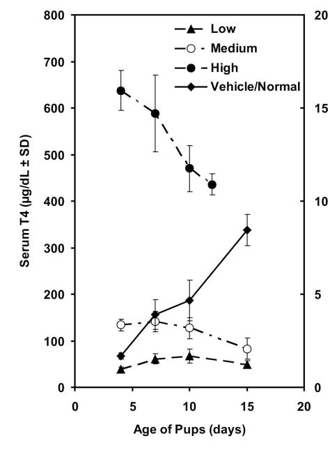
In the mature rat parotid gland, myoepithelial cells (MEC) invest intercalated ducts, but not acini. During postnatal development, however, these cells differentiate around both intercalated ducts and acini, then translocate to only intercalated ducts during weaning. Previously, we found that thyroxine (T(4)) accelerates translocation of cells with small secretory granules from acini into intercalated ducts and the number of apoptotic cells increased tremendously with high doses. We present here additional analysis of the effects of T(4) on developing rat parotid gland, namely, the distribution of MEC and the proliferation of parenchymal cells. Beginning at age four days, pups were given daily subcutaneous injections of low, medium, and high doses of T(4) or vehicle or no injection. At ages 4, 7, 10, and 15 days, glands were excised and processed for light microscopy. Sections were double-immunostained with antibodies against proliferating cell nuclear antigen (PCNA) and actin, and counterstained with hematoxylin. Proliferative activity was assessed via PCNA histochemistry and MEC were identified using actin histochemistry. MEC in the T(4) groups invested mostly acini at 15 days in vehicle/normal glands and mostly intercalated ducts after 10 days in the T(4) groups. The proliferative activity of acinar cells and MEC in vehicle/normal glands declined progressively with age and T(4) increased the rate of this decline in the MEC in a dose-dependent manner. We conclude that T(4) accelerates the translocation of MEC from acini to intercalated ducts and that an important mechanism is the more rapid decline in the proliferative activity of MEC than in acinar cells in the T(4) groups. Some of the decline in the proliferative activity of all cells in the high and medium dose T(4) groups after seven days may have been due to dose-related thyroxine toxicity.
Figures


Similar articles
-
Effects of exogenous thyroid hormone on the postnatal morphogenesis of the rat parotid gland.Anat Rec (Hoboken). 2008 Jan;291(1):94-104. doi: 10.1002/ar.20620. Anat Rec (Hoboken). 2008. PMID: 18085625
-
Differentiation of myoepithelial cells in the developing rat parotid gland.Am J Anat. 1980 Jul;158(3):299-320. doi: 10.1002/aja.1001580306. Am J Anat. 1980. PMID: 7446434
-
Secretory protein expression patterns during rat parotid gland development.Anat Rec. 1998 Nov;252(3):485-97. doi: 10.1002/(SICI)1097-0185(199811)252:3<485::AID-AR17>3.0.CO;2-J. Anat Rec. 1998. PMID: 9811227
-
Functional morphology of myoepithelial cells in the rat salivary glands: A review.J Oral Biosci. 2025 Mar;67(1):100592. doi: 10.1016/j.job.2024.100592. Epub 2024 Nov 29. J Oral Biosci. 2025. PMID: 39615670 Review.
-
Immunocytochemical studies of cell differentiation during rat salivary gland development.Eur J Morphol. 1996 Aug;34(3):149-54. doi: 10.1076/ejom.34.3.149.13032. Eur J Morphol. 1996. PMID: 8874088 Review.
Cited by
-
A perspective of comparative salivary and breast pathology. Part I: microstructural aspects, adaptations and cellular events.Eur Arch Otorhinolaryngol. 2014 Apr;271(4):647-63. doi: 10.1007/s00405-013-2488-y. Epub 2013 May 7. Eur Arch Otorhinolaryngol. 2014. PMID: 23649507 Review.
References
-
- Cher ML, Chew K, Rosenau W, Carroll PR. Cellular proliferation in prostatic adenocarcinoma as assessed by bromodeoxyuridine uptake and Ki-67 and PCNA expression. Prostate. 1995;26:87–93. - PubMed
-
- Clos J, Crepel F, Legrand C, Legrand J, Rabie A, Vigouroux E. Thyroid physiology during the postnatal period in the rat: a study of the development of thyroid function and of the morphogenetic effects of thyroxine with special reference to cerebellar maturation. Gen Com Endocr. 1974;23:178–192. - PubMed
-
- D’Agostino J, Henning SJ. Role of thyroxine in coordinate control of corticosterone and CBG in postnatal development. Am J Physiol. 1982;242:E33–E39. - PubMed
-
- Donaldson HH. Biology. In: Donaldson HH, editor. The Rat. 2. Wistar Institute; Philadelphia: 1924. pp. 15–29.
-
- Dussault JH, Coulombe P, Walker P. Effects of neonatal hyperthyroidism on the development of the hypothalamic-pituitary-thyroid axis in the rat. Endocrinology. 1982;110:1037–1042. - PubMed
Publication types
MeSH terms
Substances
Grants and funding
LinkOut - more resources
Full Text Sources
Miscellaneous