. 2009 Apr;100(4):730-6.
doi: 10.1111/j.1349-7006.2009.01098.x.

Silencing of SOCS1 in macrophages suppresses tumor development by enhancing antitumor inflammation

Affiliations

Silencing of SOCS1 in macrophages suppresses tumor development by enhancing antitumor inflammation

Masayuki Hashimoto et al. Cancer Sci. 2009 Apr.

Abstract

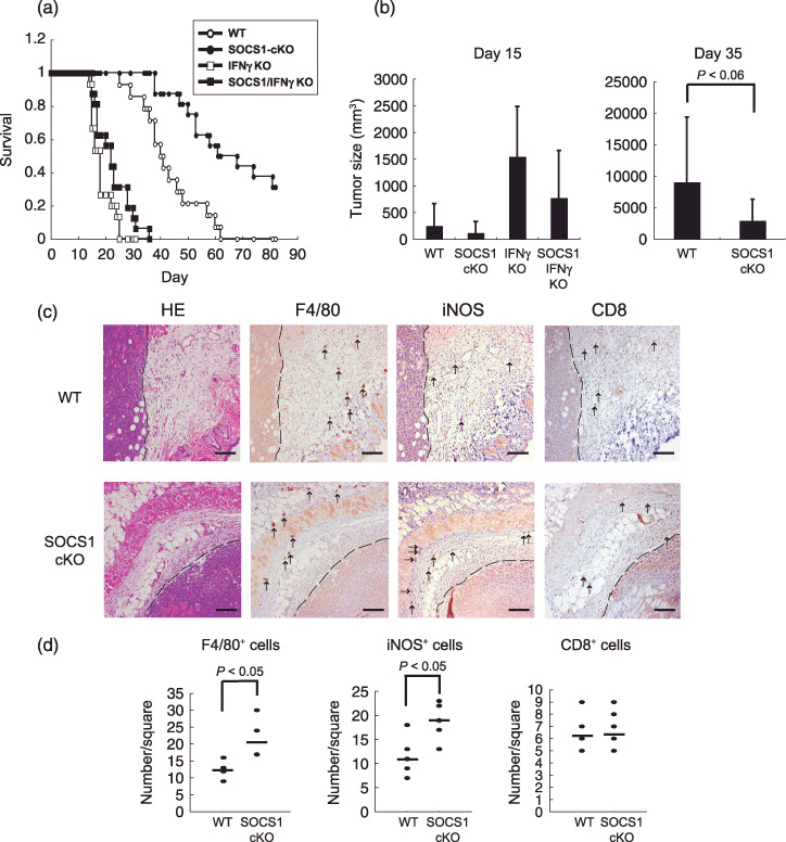
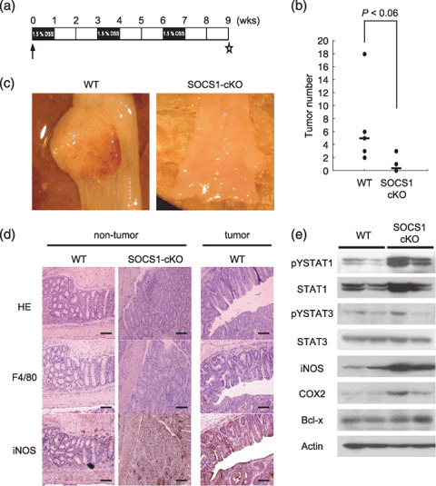
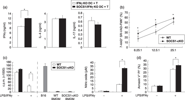
Inflammation has been shown to contribute to both tumor development and antitumor immunity. However, conditions determining these opposing effects are not well understood. Suppressor of cytokine signaling 1 (SOCS1) has been shown to play an important role in regulating inflammation and tumor development. It has been reported that silencing of SOCS1 gene in dendritic cells potentiates antitumor immunity, while SOCS1-deficiency in whole organs except for T and B cells enhances inflammation-mediated colon tumor development. To determine which types of cells are important for the suppression of tumor development by SOCS1-deficiency, we employed the conditional knockout strategy. SOCS1 gene was deleted in macrophages and neutrophils by crossing SOCS1-flox/flox mice with LysM-cre mice. Resulting conditional knockout (cKO) mice showed enhanced sensitivity to endotoxin shock. SOCS1-cKO mice survived much longer than wild-type mice after B16 melanoma transplantation. Colon carcinogenesis induced by 1,2-dimethylhydrazine (DMH) plus dextran sulfate sodium (DSS) was also reduced in SOCS1-cKO mice. SOCS1-deficiency in monocytic cells enhanced tumor-killing activity of macrophages and tumor-specific cytotoxic T cell activity. These results suggest that inflammation induced by SOCS1-deficiency in monocytes potentiates antitumor immune responses rather than tumor-promoting inflammation.


Figures

Figure 1
Figure 1
Deletion of suppressor of cytokine signaling‐1 (SOCS1) in monocytic cells in mice. (a) Quantitative real‐time reverse transcription – polymerase chain reaction analysis of SOCS1 expression level. Peritoneal macrophages were stimulated with 20 U/mL interferon (IFN)γ for 3 h. GAPDH was used as a housekeeping gene. (b) Phosphorylation of STAT1 in peritoneal macrophages in response to IFNγ. Resident peritoneal macrophages from littermate SOCS1 fl ox/fl ox wild type (WT) and LysM‐cre SOCS1 fl ox/fl ox (SOCS1‐cKO [conditional knock out]) mice were stimulated with 20 U/mL IFNγ for the indicated periods, and then cell extracts were immunoblotted with total anti‐STAT1 and antiphospho‐specific STAT1 antibody. (c) LPS sensitivity in vivo. 5‐ to 10‐week‐old SOCS1‐cKO and WT mice were intraperitoneally injected with 1.2 mg LPS (n = 11). Lethality was observed for 120 h after this challenge. The results were assessed by the logrank test. (d) Phosphorylation of STAT1, STAT3, IκBα, p65, Erk, p38, and JNK in peritoneal macrophages in response to LPS. Resident peritoneal macrophages from SOCS1‐cKO mice were stimulated with 100 ng/mL LPS for the indicated periods, and then cell extracts were immunoblotted with the indicated antibody.
Figure 2
Figure 2
Resistance of suppressor of cytokine signaling‐1 (SOCS1)‐conditional knock out (cKO) mice to B16 melanoma transplantation. (a) Wild type (WT), SOCS1‐cKO, interferon (IFN)γ−/– and SOCS1−/– IFNγ−/– mice were subcutaneously challenged with 2 × 105 B16 melanoma cells (WT: n = 14, SOCS1 LysM‐cre: n = 16, IFNγ−/– : n = 15, SOCS1−/– IFNγ−/– : n = 16). Lethality was observed for 80 days after this challenge. Kaplan‐Meier survival curves are depicted as time after tumor challenge. The results were assessed by the logrank test. P < 0.001 (b) Tumor size was assessed by measuring three perpendicular diameters of the skin tumor 15 days and 35 days after tumor challenge (WT: n = 14, SOCS1‐cKO: n = 16, IFNγ−/– : n = 8, SOCS1−/–IFNγ−/– : n = 14). (c) 3LL tumor cells (2 × 105) were injected into WT and SOCS1‐cKO mice subcutaneously. Seven days after tumor challenge, subcutaneous tumor samples were isolated and used for hematoxylin and eosin (HE) staining and immunostaining for F4/80, inducible nitric oxide synthase (iNOS) and CD8. Arrowheads show each positive cell. Bars, 50 µm. (d) The stained cell numbers in the setting square. Bar indicates the average of the number of positive cell.
Figure 3
Figure 3
Tumor formation in wild type (WT) and suppressor of cytokine signaling‐1 (SOCS1)‐conditional knock out (cKO) mice in the DSS/DMH colon cancer model. (a) Protocol of DSS/DMH. DMH (20 mg/kg body weight [b.w]) was intraperitoneally injected at indicated points. DSS (1.5%) was administrated in drinking water. Star indicates the point of sacrifice. (b) Tumor number was counted in individual mice (WT: n = 10, SOCS1‐cKO: n = 6) (c) Representative photographs of the colon from these mice in the DSS/DMH colon cancer model. (d) Hematoxylin and eosin (HE) and immunostaining for F4/80 and inducible nitric oxide synthase (iNOS). Bars, 50 µm. (e) Western blot analysis of indicated proteins in whole colonic extracts. Actin was used as a loading control.
Figure 4
Figure 4
Tumor killing activity of cytotoxic T cells (CTLs) and macrophages. (a) Cytokine production from wild type (WT) T cells activated with WT and mutant dendritic cells (DCs), bone marrow dendritic cells (BMDCs) from interferon (IFN)γ−/– or suppressor of cytokine signaling‐1 (SOCS1)−/–IFNγ−/– mice were cocultured with naïve T cells from WT mice for 72 h. Co‐cultured T cells were collected and stimulated with 1 µg/mL anti‐CD3 antibody platebound for 24 h, then IFNγ, interleukin (IL)‐4 and IL‐17 in the culture supernatant were measured by enzyme‐linked immunosorbent assay. The results are expressed as mean with SD for triplicate samples. Asterisk, P < 0.05. (b) CTL assay. Target B16 melanoma cells were injected to WT or SOCS1‐cKO (conditional knock out) mice. The spleen was taken from these mice 5 days after this challenge, and then CD8+ cells were cocultured with mitomycine C‐treated B16 cells for 5 days. CFSE‐labeled B16 target cells were cocultured with effector cells at indicated ratio for 6 h. The results are expressed as mean with SD for four independent samples. Asterisk, P < 0.05. CFSE+‐gated sulforhodamine‐valyl‐alanyl‐aspartyl‐fluoromethylketone (SR‐VAD‐FMK)+, 7 Aminoactinomycin D (AAD)+ double‐positive cells were killed by effector cells. (c) Tumor‐killing activity of macrophages. Bone marrow‐derived macrophages (BMDM) was activated with or without LPS (10 ng/mL) and IFNγ (100 U/mL). B16 cells were cocultured with BMDM from WT or SOCS1‐cKO mice for 24 h, and then B16 cell proliferation in all conditions was measured using the 3H‐thymidine uptake test (left panel). NO production was measured as nitrite using the Griess reagent (right panel). (d) Peritoneal macrophages was activated with or without LPS (10 ng/mL) and IFNγ (100 U/mL), then cocultured with B16 cells for 24 h. Apoptosis of B16 was assessed by Annexin V and propidium iodide (PI) staining. Asterisk, P < 0.001. The results are expressed as mean with SD for four independent samples.

References

    1. Sakamoto H, Yasukawa H, Masuhara M et al . A Janus kinase inhibitor, JAB, is an interferon‐gamma‐inducible gene and confers resistance to interferons. Blood 1998; 92: 1668–76. - PubMed
    1. Yasukawa H, Misawa H, Sakamoto H et al . The JAK‐binding protein JAB inhibits Janus tyrosine kinase activity through binding in the activation loop. EMBO J 1999; 18: 1309–20. - PMC - PubMed
    1. Marine JC, Topham DJ, McKay C et al . SOCS1 deficiency causes a lymphocyte‐dependent perinatal lethality. Cell 1999; 98: 609–16. - PubMed
    1. Alexander WS, Starr R, Fenner JE et al . SOCS1 is a critical inhibitor of interferon gamma signaling and prevents the potentially fatal neonatal actions of this cytokine. Cell 1999; 98: 597–608. - PubMed
    1. Tanaka K, Ichiyama K, Hashimoto M et al . Loss of suppressor of cytokine signaling 1 in helper T cells leads to defective Th17 differentiation by enhancing antagonistic effects of IFN‐gamma on STAT3 and Smads. J Immunol 2008; 180: 3746–56. - PubMed

Publication types

MeSH terms

Substances