Review
. 2009 Jun;10(6):397-409.
doi: 10.1038/nrn2647.

Neural regulation of endocrine and autonomic stress responses

Affiliations
Review

Neural regulation of endocrine and autonomic stress responses

Yvonne M Ulrich-Lai et al. Nat Rev Neurosci. 2009 Jun.

Abstract

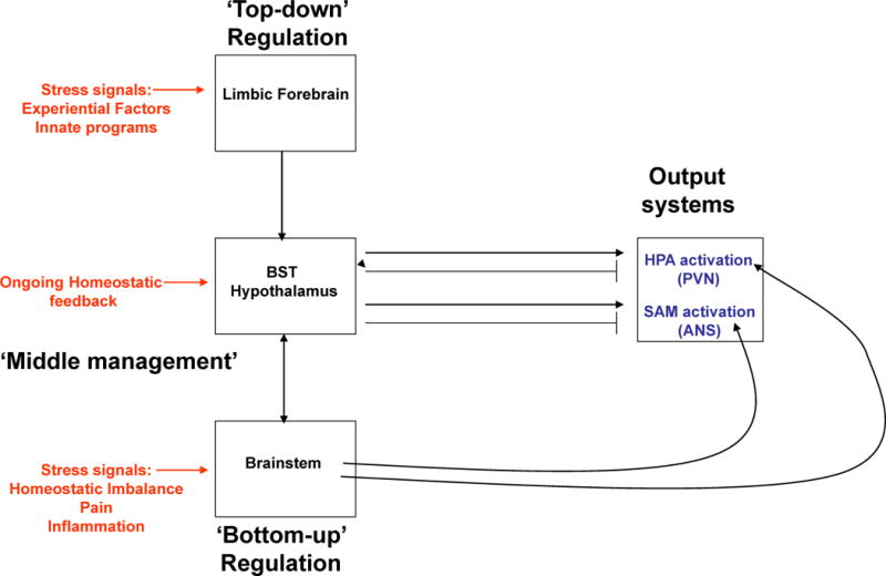
The survival and well-being of all species requires appropriate physiological responses to environmental and homeostatic challenges. The re- establishment and maintenance of homeostasis entails the coordinated activation and control of neuroendocrine and autonomic stress systems. These collective stress responses are mediated by largely overlapping circuits in the limbic forebrain, the hypothalamus and the brainstem, so that the respective contributions of the neuroendocrine and autonomic systems are tuned in accordance with stressor modality and intensity. Limbic regions that are responsible for regulating stress responses intersect with circuits that are responsible for memory and reward, providing a means to tailor the stress response with respect to prior experience and anticipated outcomes.


Figures

Figure 1
Figure 1. General scheme of brain acute stress-regulatory pathways
Stressors activate brainstem and/or forebrain limbic structures. The brainstem is able to generate rapid hypothalamus-pituitary-adrenal (HPA) axis and autonomic nervous system (ANS) responses via direct projections to hypophysiotrophic neurons in the paraventricular nucleus of the hypothalamus (PVN) or to preganglionic autonomic neurons (bottom-up regulation). In contrast, forebrain limbic regions have no direct connections with the HPA axis or the ANS and thus require intervening synapses prior to accessing autonomic or neuroendocrine neurons (top-down regulation). A high proportion of these intervening neurons are located in hypothalamic nuclei that are also responsive to homeostatic status, providing a mechanism by which the descending limbic information can be modulated according to the physiological status of the animal (middle management).
Figure 2
Figure 2. Brain circuitry regulating autonomic stress responses
Stress-induced pre-autonomic outflow originates in multiple brain areas. Colors denote brain regions that are implicated in sympathetic activation (blue), parasympathetic activation (red) or both (bicolored). The paraventricular nucleus of the hypothalamus (PVN) has substantial projections to both sympathetic and parasympathetic nuclei, including the nucleus of the solitary tract (NTS), dorsal motor nucleus of the vagus nerve (DMX), intermediolateral cell column (IML), locus coeruleus (LC) and ventrolateral medulla (VLM) (latter two not shown for clarity). The rostral VLM, LC, and PVN provide direct innervation of the IML and are thought to initiate sympathetic responses. These NTS in turn receive direct input from neurons in the infralimbic cortex (IL), central amygdala (CeA) and PVN. Other hypothalamic regions, most notably the dorsomedial hypothalamus (DMH), modulate ANS activation via connections with the PVN (and possibly other descending pathways) (see text). Parasympathetic outflow is mediated largely by descending outflow from the DMX and nucleus ambiguous (NA) (colored red) and is under the direct influence of the prelimbic cortex (PL), PVN and possibly other descending relays (see text). Parasympathetic effects of the anterior bed nucleus of the stria terminalis (aBST) are likely mediated by relays in the PVN or the NTS. The anatomical complexity of ANS integration is underscored by the mixing of sympathetic and parasympathetic projection neurons in individual nuclei.
Figure 3
Figure 3. Brain circuitry regulating HPA axis stress responses
Stress-induced activation of the dorsal part of medial parvocellular paraventricular nucleus of the hypothalamus (PVNmpd) originates in several brain regions (excitatory inputs colored blue with solid lines and inhibitory inputs (GABA) colored red with dashed lines). The paraventricular nucleus of the hypothalamus (PVN) receives direct noradrenergic, adrenergic and peptidergic innervation from the nucleus of the solitary tract (NTS). The dorsomedial component of dorsomedial hypothalamus (dmDMH) and arcuate nucleus (Arc) provide intrahypothalamic stress excitation. The anterior part of the bed nucleus of the stria terminalis (BST), particularly the anteroventral nucleus of the BST (avBST), activates HPA axis stress responses. The PVN also receives stress-excitatory drive from the avBST, dorsal raphe, tuberomammillary nucleus, supramammillary nucleus, and spinal cord, among others (omitted in the interest of space). Activation of the PVNmpd is inhibited by numerous hypothalamic circuits, including the medial preoptic area (mPOA), ventrolateral component of dorsomedial hypothalamus (vlDMH) and local neurons in the peri-PVN region (pPVN), encompassing the PVN surround and the subparaventricular zone. The posterior subregions of the bed nucleus of the stria terminalis (pBST) provides a prominent forebrain inhibition of HPA axis responses; the majority of these inputs are GABAergic.
Figure 4
Figure 4. Organization of limbic outputs
Limbic modulation of stress responses occurs predominantly via oligosynaptic inputs to the in the paraventricular nucleus of the hypothalamus (PVN) and other preautonomic brain regions. Excitatory inputs are colored blue with solid lines and inhibitory inputs (GABA) are colored red with dashed lines. Top: The ventral subiculum (vSUB) coordinates hippocampal stress output by providing glutamatergic input to primarily inhibitory PVN relays, thereby limiting psychogenic stress responses. Middle: GABAergic projections from the central amygdala (CeA) regulate responses to systemic stressors, whereas those from the medial amygdala (MeA) preferentially modulate responses to psychogenic stressors. Through glutamatergic projections within and outside the amygdala, the basolateral amygdala (BLA) plays a role in both the acute response to psychogenic stress and in chronic stress regulation. Bottom: The prelimbic cortex (PL) inhibits responses to psychogenic stress, and this inhibition is mediated predominantly by glutamatergic projections to inhibitory PVN relays. In contrast, the infralimbic cortex (IL) activates autonomic and possibly HPA axis responses to psychogenic stress, perhaps via direct (nucleus of the solitary tract, NTS) or indirect (CeA) projections. Abbreviations: anteriomedial BST (amBST), anteroventral BST (avBST), bed nucleus of the stria terminalis (BST), dorsal raphe nucleus (DRN), dorsomedial hypothalamus (DMH), lateral septum (LS), medial preoptic area (mPOA), thalamic paraventricular nucleus (PVT), peri-PVN (pPVN), posteromedial BST (pmBST), ventral subiculum (vSub).
None

References

    1. Iversen S, Iversen L, Saper CB. In: Principles of Neural Science. Kandel ER, Schwartz JH, Jessell TM, editors. Mc-Graw Hill; New York: 2000.
    1. Droste SK, et al. Corticosterone levels in the brain show a distinct ultradian rhythm but a delayed response to forced swim stress. Endocrinology. 2008;149:3244–53. - PubMed
    1. Herman JP, et al. Central mechanisms of stress integration: hierarchical circuitry controlling hypothalamo-pituitary-adrenocortical responsiveness. Front Neuroendocrinol. 2003;24:151–80. - PubMed
    1. Cunningham ET, Jr, Bohn MC, Sawchenko PE. Organization of adrenergic inputs to the paraventricular supraoptic nuclei of the hypothalamus in the rat. J Comp Neurol. 1990;292:651–67. - PubMed
    1. Cunningham ET, Jr, Sawchenko PE. Anatomical specificity of noradrenergic inputs to the paraventricular and supraoptic nuclei of the rat hypothalamus. J Comp Neurol. 1988;274:60–76. - PubMed

Publication types

MeSH terms