. 2009 Jun 24;131(24):8614-9.
doi: 10.1021/ja9014869.

Direct observation of the hole protonation state and hole localization site in DNA-oligomers

Affiliations

Direct observation of the hole protonation state and hole localization site in DNA-oligomers

Amitava Adhikary et al. J Am Chem Soc. .

Abstract

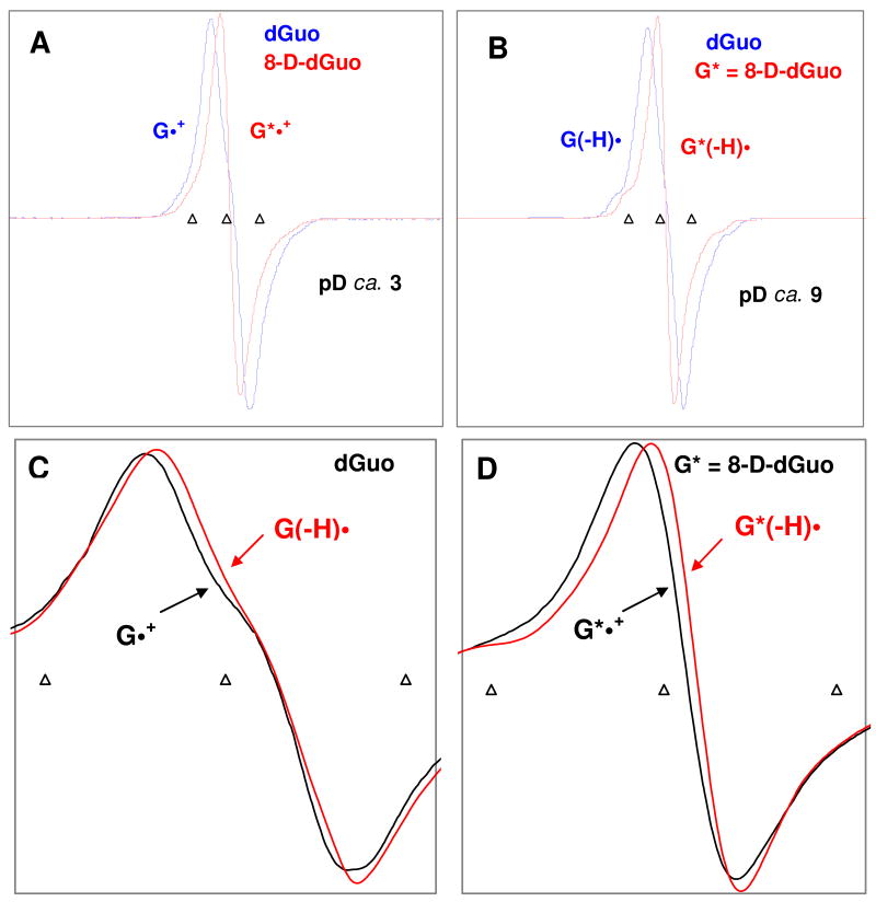
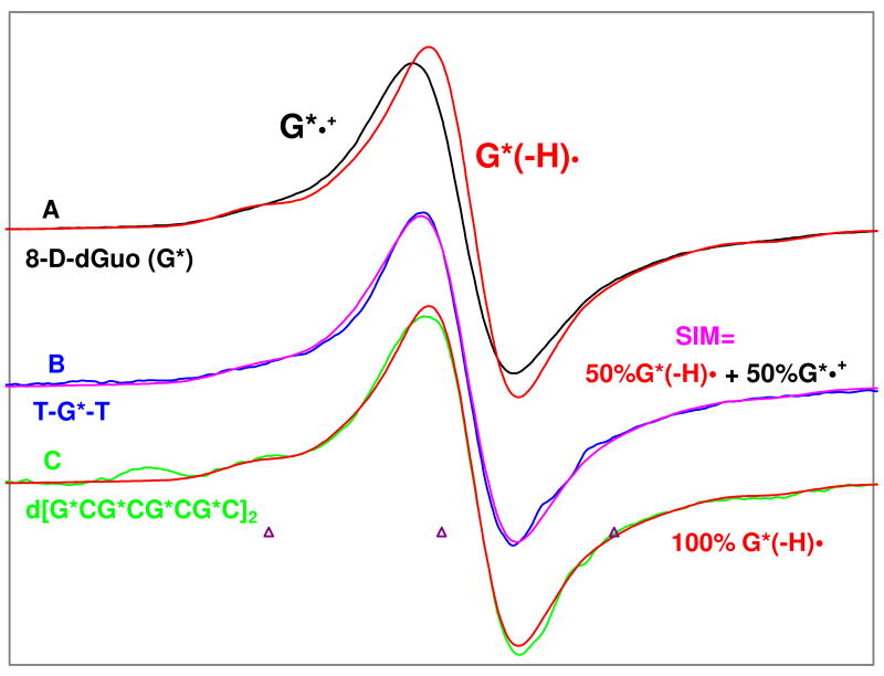
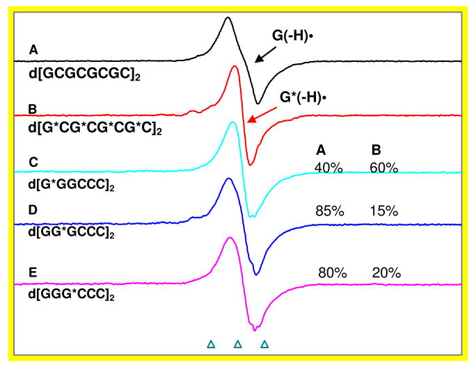
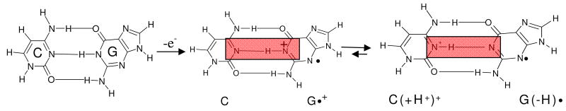
In this work, it is shown that the incorporation of an 8-deuteroguanine (G*) moiety in DNA-oligomers allows for direct determination at 77 K of (i) the location of holes (i.e., the radical site) within dsDNA at specific base sites, even within stacks of G, as well as (ii) the protonation state of the hole at that site. These findings are based on our work and demonstrate that selective deuteration at C-8 on a guanine moiety in dGuo results in an ESR signal from the guanine cation radical (G**(+)) which is easily distinguishable from that of the undeuterated guanine cation radical (G*(+)). G**(+) is also found to be easily distinguishable from its conjugate base, the N1-deprotonated radical, G*(-H)*. Our ESR results clearly establish that at 77 K (i) one-electron oxidized guanine in double stranded DNA-oligomers exists as the deprotonated neutral radical G(-H)* as a result of facile proton transfer to the hydrogen bonded cytosine and (ii) the hole is preferentially located at the 5'-end in several ds DNA-oligomers with a GGG sequence.


Figures

Figure 1
Figure 1
ESR spectra found for (A) guanine cation radicals (G•+, G*•+) and (B) deprotonated guanine neutral radicals (G(−H)•, G*(−H)•). These radicals were produced by Cl2 oxidation of dGuo (3 mg/mL) (blue) and of G* (3 mg/mL) [G* = 8-D-dGuo, 96%D] (red) at pDs ca. 3 and ca. 9 in 7.5 M LiCl glasses in D2O in the presence of K2S2O8 (5 mg/mL) as an electron scavenger. , The one-electron oxidation by Cl2- was carried out via thermal annealing at 155 K in the dark. Figures (A) and (B) clearly show that deuteration at C8, replaces the C-8-H proton hyperfine coupling from ca. 8 G in G•+ and in its N1-deprotonated neutral radical, G(−H)• with a ca. 1.2 G (C-8-D coupling) thereby narrowing the ESR spectral width and improving the spectral resolution in the deuterated species, G*•+ and G*(−H)•. Figure (C) shows that, in dGuo, G•+ (black) is not clearly distinguishable from G(−H)•; whereas Figure (D) represents that, in G*, owing to the smaller C-8-D coupling, G*•+ (black) is readily distinguishable from its N1-deprotonated neutral radical, G*(−H)• (red), because of their different lineshapes, linewidths and g⊥ values. All spectra were recorded at 77 K. The three triangular calibration markers in this figure and in the other figures represent positions of Fremy's salt resonances (the central marker is at g = 2.0056, and each of three markers is separated from one another by 13.09 G).
Figure 2
Figure 2
ESR spectra of: (A) G*•+ (black) and G*(−H)• (red) obtained from glassy (7.5 M LiCl in D2O) samples of G* [G* = 8-D-dGuo, 96% D] (3 mg/mL) , . (B) one-electron oxidized ss oligomer, TG*T, (4.5 mg/mL) in blue after subtraction of 20% of the G(−H)• spectrum (to account for 80% 8-deuteration level (see supporting information Figure S1). In Figure B, the match between the experimentally observed spectrum of one-electron oxidized TG*T (blue) with that of the simulated (pink) spectrum obtained using the spectra of G*•+ and G*(−H)• shown in (A) as benchmarks indicate that for one-electron oxidized TG*T at pD 5, G*•+ and G*(−H)• are present in equal amounts. (C) Spectrum of the one-electron oxidized ds DNA oligomer d[G*CG*CG*CG*C]2 (2 mg/mL) after subtraction of 20% of the G(−H)• spectrum (to account for 80% 8-deuteration level (see supporting information Figure S1), again with G*(−H)• from A in red superimposed. The match of green and red spectra in (C) clearly shows that, one-electron oxidized guanine in ds DNA oligomer d[G*CG*CG*CG*C]2 exists as G*(−H)•. One-electron oxidation of the monomer and the DNA-oligomers were carried out via thermal annealing at 155 K. All spectra were recorded at 77 K. Lineshape and g value differences between G*•+ and G*(− H)• allow for this analysis.
Figure 3
Figure 3
Spectra A and B are the benchmark spectra of G(−H)• and G*(−H)• (G* = 8-D-dGuo). These spectra are for the glassy samples of d[GCGCGCGC]2 and d[G*CG*CG*CG*C]2 and are assigned to G(−H)• and G*(−H)•. Note that the spectrum in B is 80% 8-deuterated. This spectrum is used as the basis function as it self corrects in our analysis. Spectra C to E are the one-electron oxidized d[GGGCCC]2 with G* moieties at 5′- end, middle and at the 3′-end of the G stack respectively. Analyses using G(−H)• and G*(−H)• spectra show that the site of hole localization is approximately 60% at the 5′-end. We have expressed the radical percentages to ±10% relative error (see materials and methods, and supporting information Figure S2). All the spectra were recorded at 77 K and the spectra of one-electron oxidized oligomers shown in Figures (A) to (E) were obtained by thermal annealing in the dark at 155 K for 20 – 30 min.
Scheme 1
Scheme 1
Schematic representation of prototropic equilibria in the G•+:C pair

References

    1. Bernhard WA. Adv Radiat Biol. 1981;9:199–280.Becker D, Sevilla MD. Adv Radiat Biol. 1993;17:121–180.Ward JF. Cold Spring Harb Symp Quant Biol. 2000;65:377–382. and references therein. von Sonntag C. Free-radical-induced DNA Damage and Its Repair. Springer-Verlag; Berlin, Heidelberg: 2006. pp. 335–447.Becker D, Adhikary A, Sevilla MD. In: Charge Migration in DNA: Physics, Chemistry and Biology Perspectives. Chakraborty T, editor. Springer-Verlag; Berlin, Heidelberg, New York: 2007. pp. 139–175.

    1. O'Neill MA, Barton JK. In: Charge Transfer in DNA: From Mechanism to Application. Wagenknecht HA, editor. Wiley-VCH Verlag GmbH & Co KGaA; Weiheim: 2005. pp. 27–75.
    1. Schuster GB, editor. Topics in Current Chemistry. Springer-Verlag; Berlin, Heidelberg: 2004. Long Range Charge Transfer in DNA. I and II.
    1. Giese B. Ann Rev Biochem. 2002;71:51–70. - PubMed
    1. Sevilla MD, Becker D, Yan M, Summerfield SR. J Phys Chem. 1991;95:3409–3415.
    2. Spalletta RA, Bernhard WA. Radiation Research. 1992;130:7–14. - PubMed
    3. Weiland B, Hüttermann J. Int J Radiat Biol. 1998;74:341–358. - PubMed
    4. Weiland B, Hüttermann J. Int J Radiat Biol. 1999;75:1169–1175. - PubMed
    5. Debije MG, Bernhard WA. J Phys Chem B. 2000;104:7845–7851.

Publication types