. 2009 May-Jun;11(3):260-8.
doi: 10.1111/j.1525-142X.2009.00328.x.

Conservation of arthropod midline netrin accumulation revealed with a cross-reactive antibody provides evidence for midline cell homology

Affiliations

Conservation of arthropod midline netrin accumulation revealed with a cross-reactive antibody provides evidence for midline cell homology

Wendy Simanton et al. Evol Dev. 2009 May-Jun.

Abstract

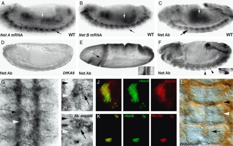
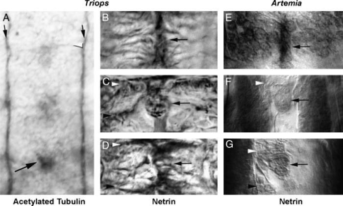
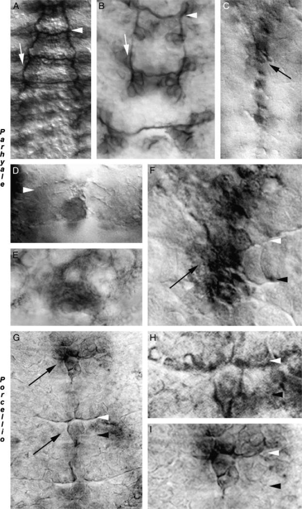
Although many similarities in arthropod CNS development exist, differences in axonogenesis and the formation of midline cells, which regulate axon growth, have been observed. For example, axon growth patterns in the ventral nerve cord of Artemia franciscana differ from that of Drosophila melanogaster. Despite such differences, conserved molecular marker expression at the midline of several arthropod species indicates that midline cells may be homologous in distantly related arthropods. However, data from additional species are needed to test this hypothesis. In this investigation, nerve cord formation and the putative homology of midline cells were examined in distantly related arthropods, including: long- and short-germ insects (D. melanogaster, Aedes aeygypti, and Tribolium castaneum), branchiopod crustaceans (A. franciscana and Triops longicauditus), and malacostracan crustaceans (Porcellio laevis and Parhyale hawaiensis). These comparative analyses were aided by a cross-reactive antibody generated against the Netrin (Net) protein, a midline cell marker and regulator of axonogenesis. The mechanism of nerve cord formation observed in Artemia is found in Triops, another branchiopod, but is not found in the other arthropods examined. Despite divergent mechanisms of midline cell formation and nerve cord development, Net accumulation is detected in a well-conserved subset of midline cells in branchiopod crustaceans, malacostracan crustaceans, and insects. Notably, the Net accumulation pattern is also conserved at the midline of the amphipod P. hawaiensis, which undergoes split germ-band development. Conserved Net accumulation patterns indicate that arthropod midline cells are homologous, and that Nets function to regulate commissure formation during CNS development of Tetraconata.


Figures

Fig. 1
Fig. 1
Anti-afrNetrin is a cross-reactive antibody. The anti-afrNet antibody was characterized in Drosophila melanogaster (fly embryos are shown in all panels except I, Aedes aegypti and L, Tribolium castaneum). Net A (A) and Net B (B) mRNA are detected in the gut (white arrows) and CNS (black arrows) of St. 14 D. melanogaster embryos. Expression is detected in these tissues with the cross-reactive anti-afrNet antibody in a slightly older embryo (C). Net protein staining is not detected in Df(1)KA9 homozygous mutant embryos (D) which lack both the NetA and NetB genes. In wild-type Drosophila embryos, the antibody recognizes staining corresponding to NetA (St. 6 cephalic fold staining marked by arrow in E is magnified at right) and NetB (lateral neuron staining marked by arrowheads in F is magnified at right) specific patterns. Furthermore, the antibody can detect ectopic Net protein expression when either NetA (J) or NetB (K) are expressed ectopically in third instar eye disc clones (ectopic expression clones marked by GFP at center, Net staining shown in red at right; overlay shown at left). These data suggest that the Net Ab recognizes Net proteins specifically, and that it can recognize both Drosophila NetA and NetB. The cross-reactive Net Ab recognizes Net protein accumulation on CNS axons (G) and midline glia in wild-type fly embryos (high magnification of single segment of St. 15 embryo is shown in H). A similar staining pattern is observed in the A. aegypti (mosquito) embryo (high magnification of midline glia in a single segment is shown in I). Comparable Net accumulation patterns are also observed in the short-germ beetle T. castaneum (marked by arrow in L; three segments shown). These data indicate that the anti-afrNet antibody is a cross-reactive antibody that specifically recognizes expression of Net proteins in multiple insects, which possess a conserved set of Net-positive midline cells. In this figure, the locations of the anterior commissures are marked with black arrowheads, while posterior commissure locations are marked with white arrowheads. Anterior is oriented left in (A–F) and up in (G–I) and (L).
Fig. 2
Fig. 2
A conserved mechanism of ventral nerve cord formation and midline Netrin accumulation in branchiopod crustaceans. Axonogenesis in Triops longicauditus was examined with antiacetylated tubulin staining (A). The temporal gradient of CNS development is evident in the 2-day-old animal shown in A, in which commissure formation (white arrowhead) is initiating in anterior segments (top), but not in the most posterior segment (bottom). In contrast, the longitudinal connectives (black arrows) have already been pioneered along the length of the entire trunk. A comparable mechanism of nerve cord formation is found in Artemia (E–G, Blanchard 1987; Duman-Scheel et al. 2007), suggesting that this mechanism for generating a nerve cord is conserved in brachiopods. The black arrow in A marks an acetylated tubulin positive cell at the midline that is observed in all of the arthropods analyzed in this investigation. Net accumulation in Triops (B–D) is detected by the cross-reactive Net Ab and resembles that of Artemia (E–G). Panels B, C, and D (Triops) are temporally comparable to panels E, F, and G, respectively, in Artemia. In branchiopods, Net accumulation is observed at the midline before commissure formation (B and E), continues as neurogenesis progresses (C, F), and is ultimately found in a subset of midline cells (D, G) that resemble those found in insects (Fig. 1). In this figure, anterior commissures are marked by white arrowheads, while posterior commissures are marked with black arrowheads in various panels; midline Net-positive cells are marked by black arrows in B–G. Anterior is oriented up in all figures.
Fig. 3
Fig. 3
Netrin accumulation in malacostracan crustaceans: detection of midline netrin accumulation in split germ band embryos. The mechanism of ventral nerve cord formation in Paryhale hawaiensis, which has a split germ band during development, was examined with anti-acetylated tubulin staining (A, B). While the anterior–posterior gradient of nerve cord formation is evident (A), the mechanism for pioneering the longitudinals observed in branchiopods is not seen in Parhyale, as evidenced by the gaps in the longitudinals (white arrows) apparent in B. Commissure formation commences despite the split germ band found in Parhyale (see lower segment in B). Net accumulation marked by the cross-reactive Ab is observed in midline cells in Parhyale (C–F). Midline Net accumulation initiates as the first commissural axons are extending (D), and is even found in split germ band segments (E), where Net-positive projections extending laterally from midline cells are observed. Net eventually accumulates in a pattern comparable to other arthropods and is detected on axons (F). Midline Net accumulation is also detected in Porcellio (G–I), another malacostracan. Higher magnification views of the middle (H) and upper (I) segments from the embryo in G are shown. In this figure, white arrowheads mark the anterior commissures, and black arrowheads mark the posterior commissures. Midline Net-positive cells are marked by black arrows. Anterior is oriented up in all figures.

Similar articles

Cited by

References

    1. Averof M, Patel NH. Crustacean appendage evolution associated with changes in Hox gene expression. Nature. 1997;388:682–686. - PubMed
    1. Blanchard CE. Pioneer neurons and the early development of the nervous system in Artemia. In: Sorgeloss P, Bengtson DA, Declair W, Jaspers E, editors. Artemia Research and its Applications. I. Universa Press; Wetteren, Belgium: 1987. pp. 5–32.
    1. Brankatschk M, Dickson BJ. Netrins guide Drosophila commissural axons at short range. Nat. Neurosci. 2006;9:188–194. - PubMed
    1. Browne WE, Price AL, Gerberding M, Patel NH. Stages of embryonic development in the amphipod crustacean, Parhyale hawaiensis. Genesis. 2005;42:124–149. - PubMed
    1. Browne WE, Schmid BGM, Wimmer EA, Martindale MQ. Expression of otd orthologs in the amphipod crustacean, Parhyale hawaiensis. Dev. Genes Evol. 2006;216:581–595. - PubMed

Publication types

LinkOut - more resources