. 2009 May 26:9:118.
doi: 10.1186/1471-2148-9-118.

Insights into the evolution of sialic acid catabolism among bacteria

Affiliations

Insights into the evolution of sialic acid catabolism among bacteria

Salvador Almagro-Moreno et al. BMC Evol Biol. .

Abstract

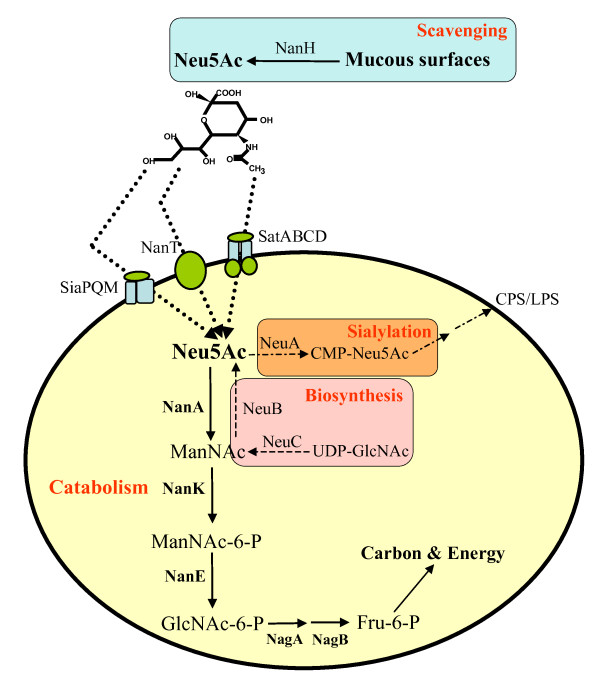
Background: Sialic acids comprise a family of nine-carbon amino sugars that are prevalent in mucus rich environments. Sialic acids from the human host are used by a number of pathogens as an energy source. Here we explore the evolution of the genes involved in the catabolism of sialic acid.

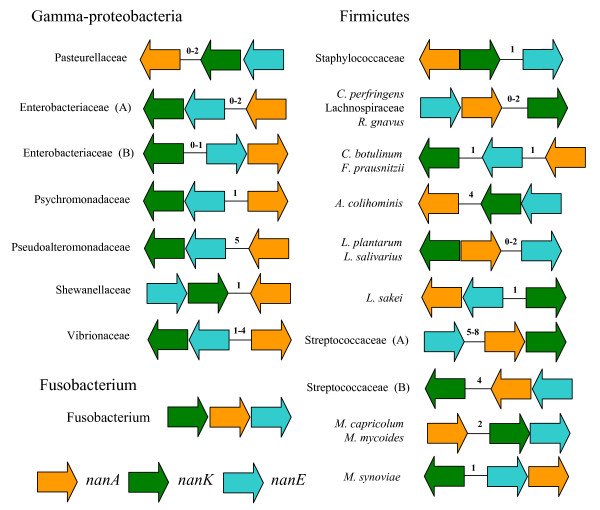
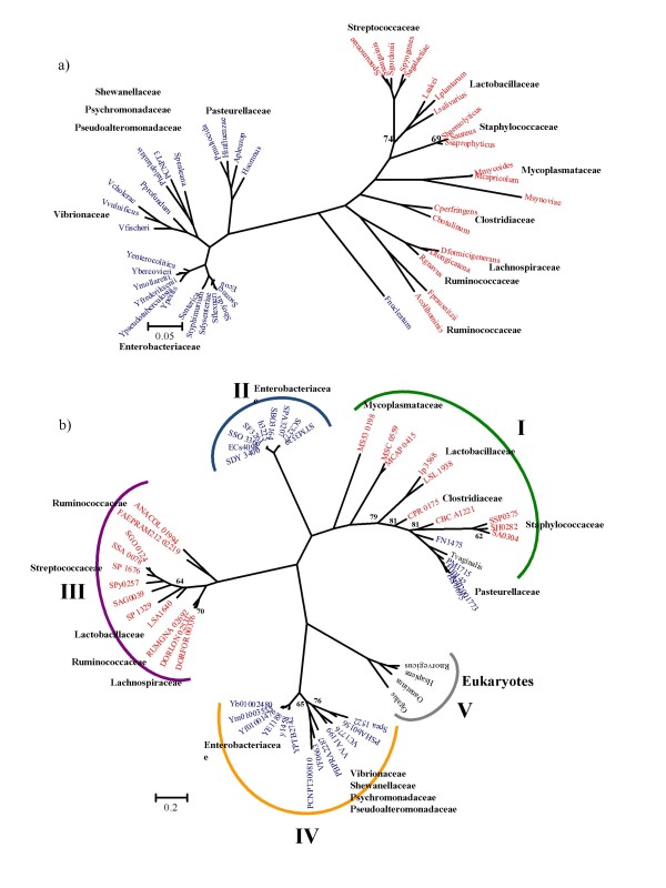
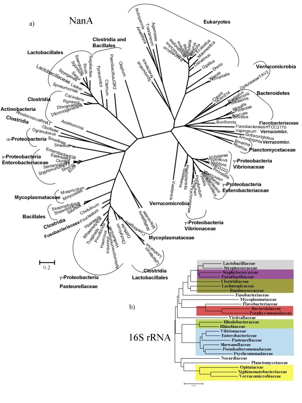
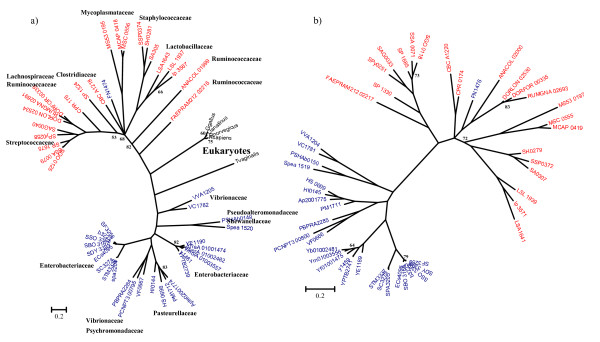
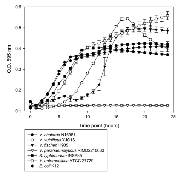
Results: The cluster of genes encoding the enzymes N-acetylneuraminate lyase (NanA), epimerase (NanE), and kinase (NanK), necessary for the catabolism of sialic acid (the Nan cluster), are confined 46 bacterial species, 42 of which colonize mammals, 33 as pathogens and 9 as gut commensals. We found a putative sialic acid transporter associated with the Nan cluster in most species. We reconstructed the phylogenetic history of the NanA, NanE, and NanK proteins from the 46 species and compared them to the species tree based on 16S rRNA. Within the NanA phylogeny, Gram-negative and Gram-positive bacteria do not form distinct clades. NanA from Yersinia and Vibrio species was most closely related to the NanA clade from eukaryotes. To examine this further, we reconstructed the phylogeny of all NanA homologues in the databases. In this analysis of 83 NanA sequences, Bacteroidetes, a human commensal group formed a distinct clade with Verrucomicrobia, and branched with the Eukaryotes and the Yersinia/Vibrio clades. We speculate that pathogens such as V. cholerae may have acquired NanA from a commensal aiding their colonization of the human gut. Both the NanE and NanK phylogenies more closely represented the species tree but numerous incidences of incongruence are noted. We confirmed the predicted function of the sialic acid catabolism cluster in members the major intestinal pathogens Salmonella enterica, Vibrio cholerae, V. vulnificus, Yersinia enterocolitica and Y. pestis.

Conclusion: The Nan cluster among bacteria is confined to human pathogens and commensals conferring them the ability to utilize a ubiquitous carbon source in mucus rich surfaces of the human body. The Nan region shows a mosaic evolution with NanA from Bacteroidetes, Vibrio and Yersinia branching closely together with NanA from eukaryotes.


Figures

Figure 1
Figure 1
Schematic representation of the metabolism of sialic acid among Bacteria. Summary of varied pathways of sialic acid utilization in Bacteria. The catabolic pathway of sialic acid involves several steps beginning with NanA. Highlighted in orange is the donor-scavenging synthesis of sialic acid. Highlighted in pink is the de novo pathway for the synthesis of sialic acid. For a more comprehensive review of sialic acid utilization see refs. 1 and 29. NanH, Neuraminidase; Neu5Ac, N-acetylneuraminic acid, sialic acid; T, sialic acid transporter; NanA, N-acetylneuraminic acid lyase; ManNAc, N-acetylmannosamine; NanK, N-acetylmannosamine kinase; ManNAc-6-P, N-acetylmannosamine-6-phosphate; NanE, N-acetylmannosamine-6-P epimerase; GlcNAc-6-P, N-acetylglucosamine-6-phosphate; NagA, N-acetylglucosamine-6-phosphate deacteylase; GlcN-6-P, Glucosamine-6-phosphate; NagB, Glucosamine-6-phosphate deaminase; Fru-6-P, Fructose-6-phosphate; NeuA, CMP-N-acetylneuraminic acid synthetase; CMP-NeuAc, CMP-N-acetylneuraminic acid; NeuB, N-acetylneuraminic acid synthase; LPS, lipopolysaccharide.
Figure 2
Figure 2
Structure of the Nan clusters among bacterial groups. Unless otherwise indicated all the members from a family shown share a common canonical structure of the Nan cluster, with differences only in the number of ORFs between the genes (indicated by a number within each cluster). nanA, orange arrows; nanK, green arrows; nanE, blue arrows. Enterobacteriaceae (A): E. coli, S. enterica, S. typhimurium, S. boydii, S. dysenteriae, S. flexneri, S. sonnei; Enterobacteriaceae (B): All species from genus Yersinia; Streptococcaceae (A) S. agalactiae, S. gordonii, S. pneumoniae (cluster A), (B) S. pyogenes, S. sanguinis, S. pneumoniae (cluster B).
Figure 3
Figure 3
Phylogenetic trees of a) 16S rRNA of bacteria containing the Nan cluster b) NanA. The trees were obtained using Bayesian analysis as implemented in MrBayes. 1,000,000 generations were used to build the consensus trees. Only confidence values below 85 are shown. Blue Operational Taxonomic Units (OTUs) indicate Gram-negative Bacteria; red OTUs, Gram-positive Bacteria; black OTUs, Eukaryotes. For NanA tree the five main lineages are highlighted with color brackets embracing them. Lineage I, green bracket; lineage II, light blue bracket; lineage III, purple bracket; lineage IV, yellow bracket; lineage V, grey bracket.
Figure 4
Figure 4
Phylogenetic trees of a) All species encoding NanA b) 16S rRNA of all families encoding NanA. The trees were obtained using Bayesian analysis as implemented in MrBayes. 1,000,000 generations were used to build the consensus trees as indicated in methods section. Main inclusive taxonomic groups are indicated. b) Grey shading, Lactobacillales; purple shading, Bacillales; dark green shading, Clostridia; red shading, Bacteroidetes; green shading, Alpha-proteobacteria; blue shading, Gamma-proteobacteria; yellow shading, Verrucomicrobia.
Figure 5
Figure 5
Phylogenetic trees of a) NanK and b) NanE. The trees were obtained using Bayesian analysis as implemented in MrBayes. 1,000,000 generations were used to build the consensus trees. Only confidence values below 85 are shown. Blue Operational Taxonomic Units (OTUs) indicate Gram-negative Bacteria; red OTUs, Gram-positive Bacteria; black OTUs, Eukaryotes. Blue branches indicate Gamma-proteobacteria; red branches, Firmicutes; Black branches; Eukaryotes.
Figure 6
Figure 6
Growth of species that encode the Nan cluster on minimal media supplemented with sialic acid. Closed circles represent V. cholerae N16961; open circles represent V. vulnificus YJO16; closed triangles represent V. fischeri H905; open triangles represent V. parahaemolyticus RIMD2210633; closed squares represent S. enterica serovar Typhimurium INSP85; open squares represent Y. enterocolitica ATCC 27729; closed diamonds represent E. coli K12.

References

    1. Vimr ER, Kalivoda KA, Deszo EL, Steenbergen SM. Diversity of microbial sialic acid metabolism. Microbiol Mol Biol Rev. 2004;68:132–153. doi: 10.1128/MMBR.68.1.132-153.2004. - DOI - PMC - PubMed
    1. Angata T, Varki A. Chemical diversity in the sialic acids and related alpha-keto acids: an evolutionary perspective. Chem Rev. 2002;102:439–469. doi: 10.1021/cr000407m. - DOI - PubMed
    1. Severi E, Hood DW, Thomas GH. Sialic acid utilization by bacterial pathogens. Microbiology. 2007;153:2817–2822. doi: 10.1099/mic.0.2007/009480-0. - DOI - PubMed
    1. Warren L. Bound Carbohydrates in Nature. Cambridge University press; 1994.
    1. Schauer R. Achievements and challenges of sialic acid research. Glycoconj J. 2000;17:485–499. doi: 10.1023/A:1011062223612. - DOI - PMC - PubMed

Publication types