Review
. 2009 Aug;137(2):466-81.
doi: 10.1053/j.gastro.2009.05.044. Epub 2009 May 24.

Stem cells and liver regeneration

Affiliations
Review

Stem cells and liver regeneration

Andrew W Duncan et al. Gastroenterology. 2009 Aug.

Abstract

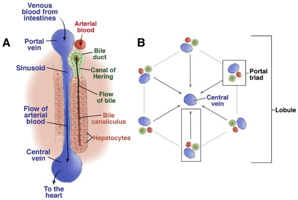
One of the defining features of the liver is the capacity to maintain a constant size despite injury. Although the precise molecular signals involved in the maintenance of liver size are not completely known, it is clear that the liver delicately balances regeneration with overgrowth. Mammals, for example, can survive surgical removal of up to 75% of the total liver mass. Within 1 week after liver resection, the total number of liver cells is restored. Moreover, liver overgrowth can be induced by a variety of signals, including hepatocyte growth factor or peroxisome proliferators; the liver quickly returns to its normal size when the proliferative signal is removed. The extent to which liver stem cells mediate liver regeneration has been hotly debated. One of the primary reasons for this controversy is the use of multiple definitions for the hepatic stem cell. Definitions for the liver stem cell include the following: (1) cells responsible for normal tissue turnover, (2) cells that give rise to regeneration after partial hepatectomy, (3) cells responsible for progenitor-dependent regeneration, (4) cells that produce hepatocyte and bile duct epithelial phenotypes in vitro, and (5) transplantable liver-repopulating cells. This review will consider liver stem cells in the context of each definition.


Figures

Figure 1
Figure 1
Structure of the hepatic lobule. (A) The portal triad consists of bile ducts, hepatic artery, and portal vein. Mixed blood from the hepatic artery and portal vein flows past hepatocytes through the sinusoids, covered with fenestrated endothelial cells to the central vein. Bile produced by the hepatocytes is collected in the bile canaliculus and flows towards the bile duct. The Canal of Hering is the junction between the hepatic plate and the bile ducts. This is the region where oval cell precursors reside. (B) Each hepatic lobule consists of 1 central vein and 6 surrounding portal triads.
Figure 2
Figure 2
Progenitor lineage relationships in adult liver and pancreas. (A) Classic model depicting one single hepatic oval cell type, which is the immediate off-spring of the intrahepatic oval cell progenitor and immediate precursor to both hepatocytes and bile ducts. Dashed lines delineate rare or hypothetical cell fate transitions that occur only under specific experimental conditions. (B) Oval cell heterogeneity model. Different stages of oval cell maturation exist, differing in proliferative potential as well as gene expression. The most mature oval cell is bipotential and gives rise to pre-hepatocytes and pre-ductal cells, which are not yet fully mature. (C) Unilineage model. Distinct unilineage progenitor cells give rise to bile ducts or hepatocytes, possibly by differentiating through intermediate cell types. These progenitor cells could comprise the oval cell population.
Figure 3
Figure 3
Signaling events during the hepatic oval cell response. A time line representing the stages of oval cell activation: activation, proliferation, migration, and differentiation. The factors that are involved in each stage of the response are listed at the bottom.
Figure 4
Figure 4
Cells with adult liver-repopulating potential. (A) Unipotential liver-repopulating cells. Hepatocytes and bile duct epithelial cells regenerate during normal tissue turnover. Under defined experimental conditions, pancreatic progenitor cells and MSCs differentiate into hepatocytes, and HSC-derived myelomonocytic cells fuse with hepatocytes. (B) Bipotential liver-repopulating cells. Intrahepatic liver stem cells and/or oval cells differentiate into hepatocytes and bile duct epithelial cells (ie, oval cell response). Fetal hepatoblasts, derived from pluripotent ESCs, also differentiate into hepatocytes and bile duct epithelial cells in transplantation experiments.

References

    1. Desmet VJ. Organizational principles. In: Arias IM, editor. The liver—biology and pathobiology. Raven; New York, NY: 1994. pp. 3–14.
    1. Mall FP. A study of the structural unit of the liver. Am J Anat. 1906;5:227–308.
    1. Wisse E. An electron microscopic study of the fenestrated endothelial lining of rat liver sinusoids. J Ultrastruct Res. 1970;31:125–150. - PubMed
    1. Jungermann K, Kietzmann T. Zonation of parenchymal and non-parenchymal metabolism in liver. Annu Rev Nutr. 1996;16:179–203. - PubMed
    1. Moorman AF, Vermeulen JL, Charles R, et al. Localization of ammonia-metabolizing enzymes in human liver: ontogenesis of heterogeneity. Hepatology. 1989;9:367–372. - PubMed

Publication types

MeSH terms