. 2009 Nov;50(11):2314-23.
doi: 10.1194/jlr.P900032-JLR200. Epub 2009 May 24.

Myocardial lipid accumulation in patients with pressure-overloaded heart and metabolic syndrome

Affiliations

Myocardial lipid accumulation in patients with pressure-overloaded heart and metabolic syndrome

Raffaele Marfella et al. J Lipid Res. 2009 Nov.

Abstract

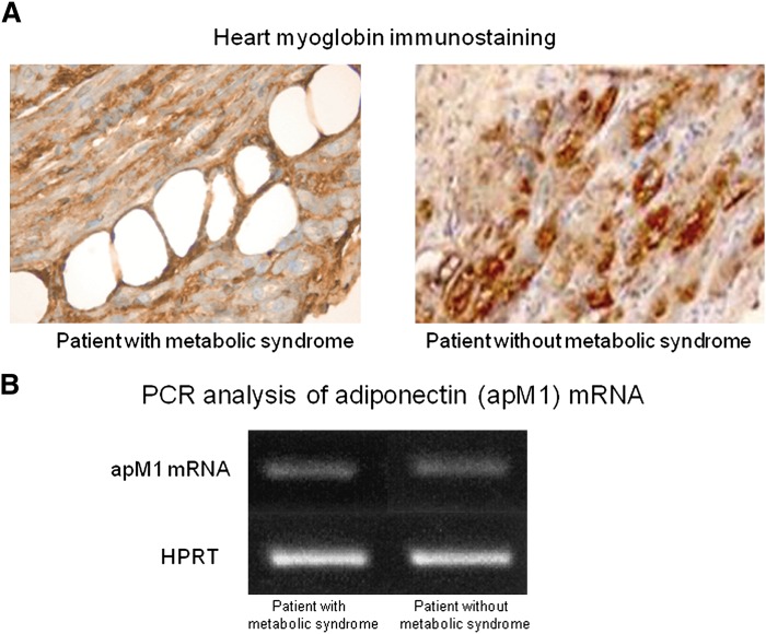
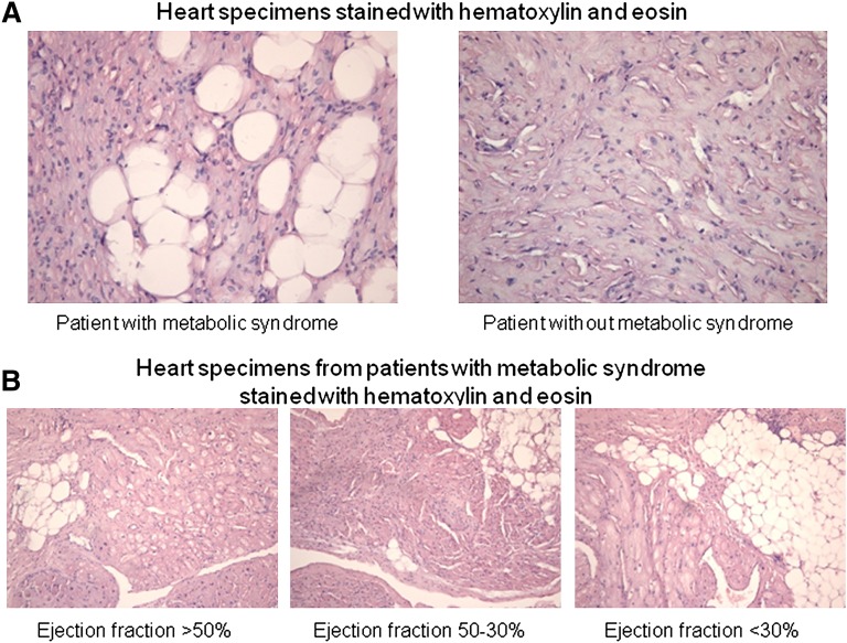
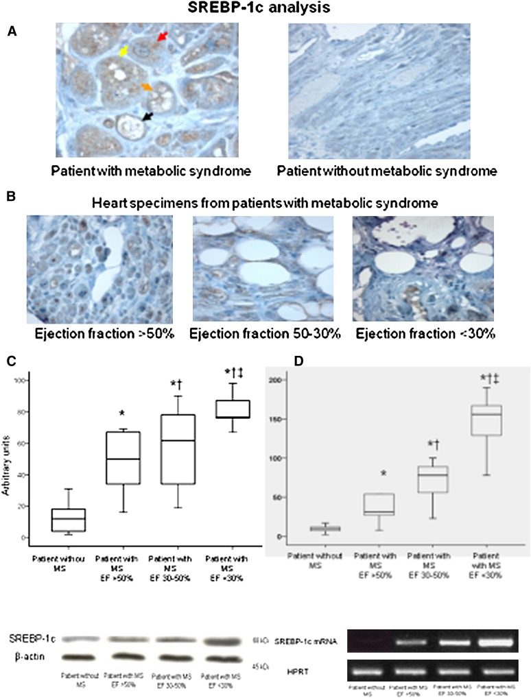
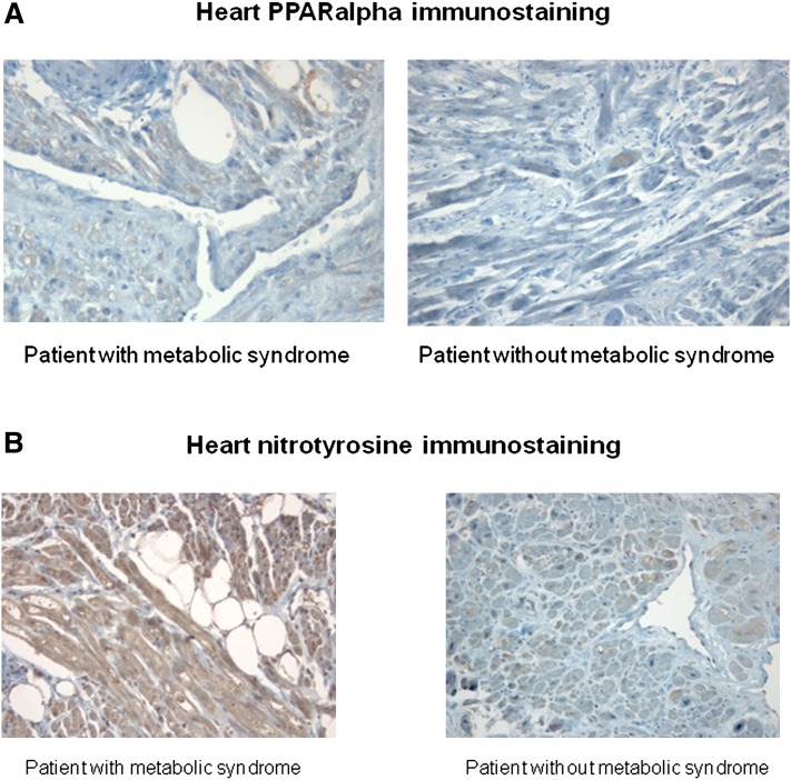
We evaluated the role of sterol-regulatory element binding protein (SREBP)-1c/peroxisome proliferator activated receptor-gamma (PPARgamma) pathway on heart lipotoxicity in patients with metabolic syndrome (MS) and aortic stenosis (AS). Echocardiographic parameters of heart function and structural alterations of LV specimens were studied in patients with (n = 56) and without (n = 61) MS undergoing aortic valve replacement. Tissues were stained with hematoxylin-eosin (H and E) and oil red O for evidence of intramyocyte lipid accumulation. The specimens were also analyzed with PCR, Western blot, and immunohistochemical analysis for SREBP-1c and PPARgamma. Ejection fraction (EF) was lower in MS compared with patients without MS (P < 0.001); no difference was found in aortic orifice surface among the groups. H and E and oil red O staining of specimens from MS patients revealed several myocytes with intracellular accumulation of lipid, whereas these alterations were not detected in biopsies from patients without MS. Patients without MS have low levels and weak immunostaining of SREBP-1c and PPARgamma in heart specimens. In contrast, strong immunostaining and higher levels of SREBP-1c and PPARgamma were seen in biopsies from the MS patients. Moreover, we evidenced a significative correlation between both SREBP-1c and PPARgamma and EF and intramyocyte lipid accumulation (P < 0.001). SREBP-1c may contribute to heart dysfunction by promoting lipid accumulation within myocytes in MS patients with AS; SREBP-1c may do it by increasing the levels of PPARgamma protein.


Figures

Fig. 1.
Fig. 1.
Representative immunohistochemical analysis of myoglobin protein from ventricular biopsy specimens (×600). The specimens from both patients with and without metabolic syndrome show a strong immunostaining for myoglobin protein in hypertrophied myocytes (A). Representative PCR analysis of apM1 mRNA content in heart specimens from patients without metabolic syndrome: apM1 transcripts were almost undetectable in heart specimens of patients with and without metabolic syndrome (B).
Fig. 2.
Fig. 2.
Vacuolated myocytes in representative ventricular-biopsy specimens hematoxylin and eosin. The specimen from a patient without metabolic syndrome shows hypertrophied myocytes without vacuoles (×600). The specimens from a patient with metabolic syndrome shows a high number of vacuolated myocytes (×600) (A). Specimens from patients with metabolic syndrome show a progressive increase in vacuolated myocytes according to the ejection fraction (×400) (B).
Fig. 3.
Fig. 3.
Representative immunohistochemical analysis of SREBP-1c protein from ventricular biopsy specimens (×600). The specimen from a patient without metabolic syndrome shows a weak immunostaining for SREBP-1c protein in hypertrophied myocytes. The specimen from a patient with metabolic syndrome shows a strong immunostaining in hypertrophied myocytes as well as in vacuolated myocytes in which the staining is localized around the vacuoles. In addition, we observe several stages representative for the different progression from intense staining to completely vacuolated myocyte in the same specimen: in the section analyzed the specimen shows a hypertrophied myocyte with a strong immunostaining without vacuoles (red arrow); yellow arrow evidences a few vacuoles in the myocyte with a strong immunostaining; orange arrow indicates numerous vacuoles in the myocytes; at this stage the vacuoles are surrounded by intense staining in the cytoplasm; finally, at this stage, probably, the vacuoles flow together in a large vacuole that occupies the cytoplasm completely (black arrow) (A). Specimens from patients with metabolic syndrome show a progressive increase in both vacuolated myocytes and immureactivity for SREBP-1c protein according to the ejection fraction (×400) (B). Western blot analysis of SREBP-1c protein contents in heart specimens from patients with metabolic syndrome according to the ejection fraction. P < 0.05 versus patients with ejection fraction between 30% and 50%. P < 0.05 versus patients with ejection fraction >50%. * P < 0.05 versus patients without metabolic syndrome. Representative western blot analysis of SREBP-1c protein content in heart specimens from patients with metabolic syndrome according to the ejection fraction. C: Box plot showing SREBP-1 mRNA levels in ventricular biopsies from patients without metabolic syndrome and from patients with metabolic syndrome according to ejection fraction. The mRNA levels were measured with RT-PCR. Representative PCR analysis of SREBP-1c mRNA content in heart specimens from patients without metabolic syndrome and from patients with metabolic syndrome according to the ejection fraction (D).
Fig. 4.
Fig. 4.
Representative immunohistochemical analysis of PPARγ protein from ventricular biopsy specimens (×400). The specimen from a patient without metabolic syndrome did not show immunostaining for PPARγ protein in hypertrophied myocytes. The specimen from a patient with metabolic syndrome shows a strong immunostaining for PPARγ protein in hypertrophied myocytes as well as in vacuolated myocytes in which the staining is localized around the vacuoles (A). Specimens from patients with metabolic syndrome show a progressive increase in both vacuolated myocytes and immureactivity for PPARγ protein according to the ejection fraction (×400) (B). Western blot analysis of PPARγ protein contents in heart specimens from patients with metabolic syndrome according to the ejection fraction. P < 0.05 versus patients with ejection fraction between 30% and 50%. P < 0.05 versus patients with ejection fraction >50%. * P < 0.05 versus patients without metabolic syndrome. Representative western blot analysis of PPARγ protein content in heart specimens from patients with metabolic syndrome according to the ejection fraction (C). Box plot showing PPARγ mRNA levels in ventricular biopsies from patients without metabolic syndrome and from patients with metabolic syndrome according to ejection fraction. The mRNA levels were measured with RT-PCR. Representative PCR analysis of PPARγ mRNA content in heart specimens from patients without metabolic syndrome and from patients with metabolic syndrome according to the ejection fraction (D).
Fig. 5.
Fig. 5.
Representative immunohistochemical analysis of PPARα protein from ventricular biopsy specimens (×400). The specimens from patients with and without metabolic syndrome show a similar immunostaining for PPARα protein in myocytes (A). Representative immunohistochemical analysis of nitrotyrosine from ventricular biopsy specimens (×200). The specimen from patients with metabolic syndrome shows a strong immunostaining for nitrotyrosine in hypertrophied myocytes (B).

Comment in

References

    1. Burchfiel C. M., Skelton T. N., Andrew M. E., Garrison R. J., Arnett D. K., Jones D. W., Taylor H. A., Jr 2005. Metabolic syndrome and echocardiographic left ventricular mass in blacks: the Atherosclerosis Risk in Communities (ARIC) Study. Circulation. 112: 819–827. - PubMed
    1. McGavock J. M., Victor R. G., Unger R. H., Szczepaniak L. S. 2006. Adiposity of the heart, revisited. Ann. Intern. Med. 144: 517–524. - PubMed
    1. McGavock J. M., Lingvay I., Zib I., Tillery T., Salas N., Unger R., Levine B. D., Raskin P., Victor R. G., Szczepaniak L. S. 2007. Cardiac steatosis in diabetes mellitus: a 1H-magnetic resonance spectroscopy study. Circulation. 116: 1170–1175. - PubMed
    1. Zhou Y. T., Grayburn P., Karim A., Shimabukuro M., Higa M., Baetens D., Orci L., Unger R. H. 2000. Lipotoxic heart disease in obese rats: implications for human obesity. Proc. Natl. Acad. Sci. USA. 97: 1784–1789. - PMC - PubMed
    1. Chiu H. C., Kovacs A., Ford D. A., Hsu F. F., Garcia R., Herrero P. 2001. A novel mouse model of lipotoxic cardiomyopathy. J. Clin. Invest. 107: 813–822. - PMC - PubMed

MeSH terms