. 2009 Jun 9;106(23):9197-202.
doi: 10.1073/pnas.0900153106. Epub 2009 May 22.

Direct functional interaction of initiation factor eIF4G with type 1 internal ribosomal entry sites

Affiliations

Direct functional interaction of initiation factor eIF4G with type 1 internal ribosomal entry sites

Sylvain de Breyne et al. Proc Natl Acad Sci U S A. .

Abstract

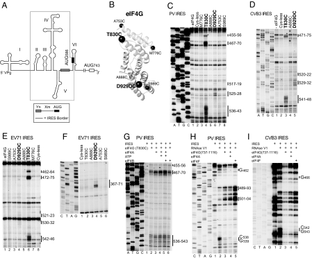
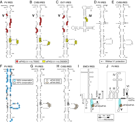
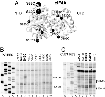
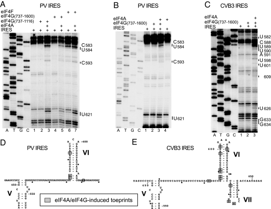
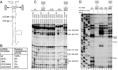
Viral internal ribosomal entry sites (IRESs) mediate end-independent translation initiation. There are 4 major structurally-distinct IRES groups: type 1 (e.g., poliovirus) and type 2 (e.g., encephalomyocarditis virus), which are dissimilar except for a Yn-Xm-AUG motif at their 3' borders, type 3 (e.g., hepatitis C virus), and type 4 (dicistroviruses). Type 2-4 IRESs mediate initiation by distinct mechanisms that are nevertheless all based on specific noncanonical interactions with canonical components of the translation apparatus, such as eukaryotic initiation factor (eIF) 4G (type 2), 40S ribosomal subunits (types 3 and 4), and eIF3 (type 3). The mechanism of initiation on type 1 IRESs is unknown. We now report that domain V of type 1 IRESs, which is adjacent to the Yn-Xm-AUG motif, specifically interacts with the central domain of eIF4G. The position and orientation of eIF4G relative to the Yn-Xm-AUG motif is analogous in type 1 and 2 IRESs. eIF4G promotes recruitment of eIF4A to type 1 IRESs, and together, eIF4G and eIF4A induce conformational changes at their 3' borders. The ability of mutant type 1 IRESs to bind eIF4G/eIF4A correlated with their translational activity. These characteristics parallel the mechanism of initiation on type 2 IRESs, in which the key event is binding of eIF4G to the J-K domain adjacent to the Yn-Xm-AUG motif, which is enhanced by eIF4A. These data suggest that fundamental aspects of the mechanisms of initiation on these unrelated classes of IRESs are similar.


Conflict of interest statement

The authors declare no conflict of interest.

Figures

Fig. 1.
Fig. 1.
Specific binding of eIF4G to type 1 IRESs identified by directed hydroxyl radical cleavage and enzymatic footprinting. (A) Model of the PV1M 5′ UTR, showing its type 1 IRES (boxed), the Yn-Xm-AUG motif, the initiation codon AUG743, and domains I–VI. (B) Model of eIF4G's central domain (Protein Data Bank ID code 1HU3), with spheres labeled to show the positions of cysteines used to tether Fe(II)-BABE. (C–G) Primer extension analysis of directed hydroxyl radical cleavage of PV (C and G), CVB3 (D), and EV71 (E and F) IRESs from Fe(II)-tethered eIF4G in IRES/eIF4G complexes in the absence (C–E) and presence of eIF4A/eIF4B (G), as indicated. (H and I) Primer extension analysis of residues in PV (H) and CVB3 (I) IRESs protected from RNase V1 cleavage by eIF4G, eIF4G/eIF4A, and eIF4F, as indicated. Sites of hydroxyl radical cleavage and residues with altered sensitivity to RNase V1 cleavage are indicated to the right. Lanes A, T, G, and C depict sequences generated from the same primers.
Fig. 2.
Fig. 2.
Sites of interaction of elF4G and elF4A with CVB3, EV71, and PV Type 1 IRESs. (A–C) Sites of directed hydroxyl radical cleavage from positions 830 and 929 of eIF4G737–1116 mapped onto PV Domain V (A), CVB3 domain V (B), and EV71 domains IV and V (C). (D and E) Nucleotides protected by eIF4G from RNase V1 cleavage mapped onto PV (D) and CVB3 domain V (E). (F) Sequence conservation in domain V of enteroviruses. (G and H) Sites of directed hydroxyl radical cleavage from Cys-33 and Cys-42 of eIF4A mapped onto PV (G) and CVB3 domain V (H). (I and J) Models of the type 2 EMCV IRES (I) and the type 1 PV IRES (J), showing the proximity and orientation of the bound eIF4G/eIF4A complex relative to the Yn-Xm-AUG motif.
Fig. 3.
Fig. 3.
Recruitment of eIF4A to type 1 IRESs by eIF4G. (A) Model of eIF4A (Protein Data Bank ID code 3EX7), with spheres labeled to show the positions of cysteines used to tether Fe(II)-BABE. (B and C) Primer extension analysis of directed hydroxyl radical cleavage of PV (B) and CVB3 (C) IRESs from Fe(II)-tethered eIF4A in IRES/eIF4G/eIF4A complexes. Sites of hydroxyl radical cleavage are indicated to the right. Lanes A, T, G, and C depict sequences generated from the same primers.
Fig. 4.
Fig. 4.
Conformational changes induced by eIF4G/4A at the 3′ border of PV and CVB3 IRESs. (A–C) Toe-printing analysis was done on PV (A and B) and CVB3 (C) IRESs in the presence of ATP and eIFs as indicated. Lanes A, T, G, and C depict sequences generated from the same primers. The positions of stops caused by the presence of eIFs are indicated to the right. (D and E) The positions of stops are mapped onto PV domain VI (D) and CVB3 domains VI and VII (E) (25, 26).
Fig. 5.
Fig. 5.
Determinants of binding of eIF4G/eIF4A to domain V of the PV IRES. (A and B) The effects of mutations in PV domain V (A) were assayed by translation in vitro (B). Values represent the translational activity of IRES mutants as a percentage of the value for the WT IRES, and are the mean of 3 assays. (C) Primer extension analysis of directed hydroxyl radical cleavage of PV IRES mutants in eIF4G/IRES complexes by Fe(II)-tethered eIF4G737–1116(Cys-830), and in eIF4G/eIF4A/IRES complexes by Fe(II)-tethered eIF4A-Cys-33 and eIF4A-Cys-42, as indicated. (D) Toe-printing analysis of conformational changes induced by eIF4G/4A at the 3′ border of PV IRES mutants. Lanes A, T, G, and C depict sequences generated from the same primer.

References

    1. Pestova TV, Lorsch J, Hellen CUT. In: Translational Control in Biology and Medicine. Mathews M, Sonenberg N, Hershey J, editors. Cold Spring Harbor, NY: Cold Spring Harbor Lab Press; 2007. pp. 87–120.
    1. Pestova TV, Shatsky IN, Fletcher SP, Jackson RJ, Hellen CUT. A prokaryotic-like mode of cytoplasmic eukaryotic ribosome binding to the initiation codon during internal translation initiation of hepatitis C and classical swine fever virus RNAs. Genes Dev. 1998;12:67–83. - PMC - PubMed
    1. Wilson JE, Pestova TV, Hellen CU, Sarnow P. Initiation of protein synthesis from the A site of the ribosome. Cell. 2000;102:511–520. - PubMed
    1. Pestova TV, Hellen CUT, Shatsky IN. Canonical eukaryotic initiation factors determine initiation of translation by internal ribosomal entry. Mol Cell Biol. 1996;16:6859–6869. - PMC - PubMed
    1. Lomakin IB, Hellen CU, Pestova TV. Physical association of eukaryotic initiation factor 4G (eIF4G) with eIF4A strongly enhances binding of eIF4G to the internal ribosomal entry site of encephalomyocarditis virus and is required for internal initiation of translation. Mol Cell Biol. 2000;20:6019–6029. - PMC - PubMed

Publication types

MeSH terms

LinkOut - more resources