. 2009 Jun 9;106(23):9157-62.
doi: 10.1073/pnas.0901240106. Epub 2009 May 22.

Structure of toroidal DNA collapsed inside the phage capsid

Affiliations

Structure of toroidal DNA collapsed inside the phage capsid

Amélie Leforestier et al. Proc Natl Acad Sci U S A. .

Abstract

The structure of DNA toroids made of individual DNA molecules of various lengths (3,000 to 55,000 bp) was studied, by using partially filled bacteriophage capsids in conjunction with cryoelectron microscopy. The tetravalent cation spermine was diffused through the capsid to condense the DNA under conditions that were chosen to produce a hexagonal packing. Our results demonstrate that the frustration arising between chirality and hexagonal packing leads to the formation of twist walls; the correlation between helices combined with their strong curvature impose variations of the DNA helical pitch.


Conflict of interest statement

The authors declare no conflict of interest.

Figures

Fig. 1.
Fig. 1.
Cryoelectron microscopy of T5 bacteriophages after partial DNA ejection under pressure and spermine-induced condensation of the DNA kept in the capsid. (A) Full and empty capsids coexist with capsids containing intermediate amounts of DNA. In this latter situation, DNA forms toroids or related shapes that are seen in top view (1), side view (2) and oblique views (3). (B) Micrographs taken at a larger defocus let us recognize the bacteriophage tail connected at one apex of the capsid, but prevent the observation of the details of the toroidal DNA structure. (C–H) Projections of the capsids along their 5-fold axis (C and D), 2-fold axis (E and F), and 3-fold axis (G and H) present different views of the icosahedral capsid. Side (C, E, and G) and top views (D, F, and H) of the toroids can be found for each of these characteristic projection views. The arrow in G underlines the alignments of DNA parallel to the capsid surface. (Scale bar, 50 nm.)
Fig. 2.
Fig. 2.
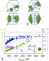
Schematic representations of side view toroids of different sizes. (A–D) Small (A), medium (B and C), and large (D), with the definition of the inner (rin) and outer radii (rout) measured in each situation to calculate LDNAin. (E) The outer radius rout, the R/r ratio and the surface S of the cross-section are expressed as a function of LDNA. The error in the measurements is estimated to vary from 5% in large toroids to 15% in the smallest ones.
Fig. 3.
Fig. 3.
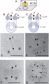
Patterns observed (D–G) and expected (A–C) in top and side views of the toroids. The z axis of the toroid may be aligned parallel to T2 (B and D), parallel to θ2 (C and E), parallel to T2 on one side and to θ2 on the other side (F), or to others directions of the hexagonal lattice. Lateral projections of the lattice should reveal periodic striations separated by ah/2 or ah√3/2, with ah the interhelix spacing, depending on the plane of projection relative to the directions of the hexagonal lattice. Only ah√3/2 striations are seen, on top and side views of the toroids (arrows), and interrupted by regions where striations are absent or unclear (*) (D–G). (Scale bar, 50 nm.)
Fig. 4.
Fig. 4.
Bundles of hexagonally packed DNA helices observed outside of the capsids. (A–C) Bundles show an alternation of domains with (arrow) and without (*) visible striated patterns revealing the presence of twist walls. In striated domains, the pitch P = 3.3 nm and spacing d = 2.5 nm can be observed directly on the micrographs (B and C) and measured from the Fourier transform (D) by comparison with the TMV virus reference (arrowhead at 1/2.3 nm in C and D). (E) Helices are phased as sketched in top and side views. In top view, the small and large grooves of the double helix are drawn in blue and red respectively. (Scale bars, 20 nm.)
Fig. 5.
Fig. 5.
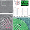
DNA correlations and change of the pitch of the double helices in the toroidal structure. (A and B) Digitized and filtered image of the toroid displayed in Fig. 3G, highlighting the details of the striated domains, with the DNA helical pitch P and spacing d. (C) The periodic contrast between helices arises from the DNA alignment, with a P/2 shift between 2 adjacent rows as sketched on top and side view. (D) The variation of the DNA helical pitch P along a given row of the toroid. (E) For comparison, the unconfined large toroid reveals the same phasing of DNA helices. (Scale bar, 20 nm.) [E is reproduced with permission from refs. (Copyright 2001, National Academy of Sciences) and (Copyright 2008, BioMed Central).]
Fig. 6.
Fig. 6.
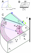
Toroidal model. (A–C) From the analysis of 2 toroids whose dimensions overlap partially (A), we propose a sketch of the structure along 2 orthogonal planes P1 and P2, respectively parallel and normal to the z axis of the toroid (B and C). Only 1 period is drawn that corresponds to 1 striated domain (pink) and 1 unresolved domain (blue) with a twist wall in between that may extend into the blue zone. The DNA hexagonal lattice of parameter ah (in P1) is seen as a series of stripes separated by d = ah√3/2 in P2. DNA helices are bent and correlated, which defines a second hexagonal lattice in P2 (ah = P), with P the DNA helical pitch, which varies over a period. Dislocation lines (L) correspond to the introduction of additional DNA helical pitches. Their density and distribution are not known.

References

    1. Gosule LC, Schellman JA. Compact form of DNA induced by spermidine. Nature. 1976;259:333–335. - PubMed
    1. Laemmli UK. Characterization of DNA condensates induced by poly (ethylene oxide) and polylysine. Proc Natl Acad Sci USA. 1975;72:4288–4292. - PMC - PubMed
    1. Chattoraj DK, Gosule LC, Schellman A. DNA condensation with polyamines. II. Electron microscopic studies. J Mol Biol. 1978;121:327–337. - PubMed
    1. Widom J, Baldwin RL. Cation-induced toroidal condensation of DNA studies with Co3+(NH3) 6. J Mol Biol. 1980;144:431–453. - PubMed
    1. Lerman L. A transition to a compact form of DNA in polymer solutions. Proc Natl Acad Sci USA. 1971;68:1886–1890. - PMC - PubMed

Publication types

MeSH terms

LinkOut - more resources