. 2009 Jun 15;23(12):1423-37.
doi: 10.1101/gad.1805709. Epub 2009 May 26.

AdnAB: a new DSB-resecting motor-nuclease from mycobacteria

Affiliations

AdnAB: a new DSB-resecting motor-nuclease from mycobacteria

Krishna Murari Sinha et al. Genes Dev. .

Abstract

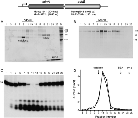
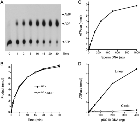
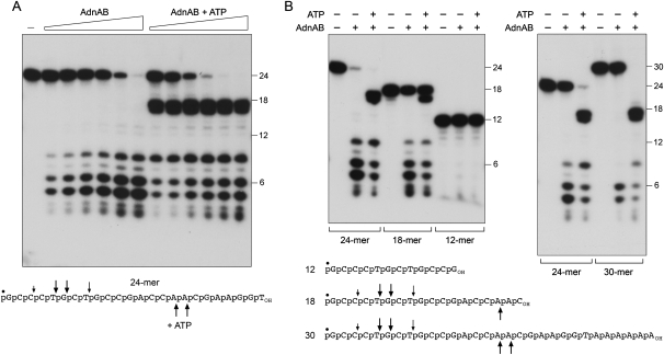
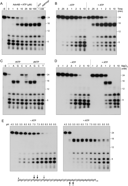
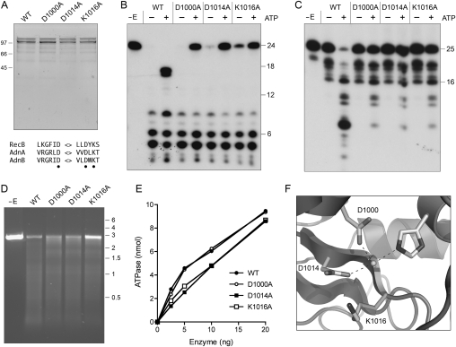
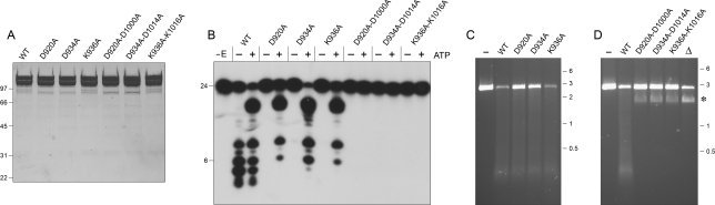
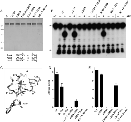
The resection of DNA double-strand breaks (DSBs) in bacteria is a motor-driven process performed by a multisubunit helicase-nuclease complex: either an Escherichia coli-type RecBCD enzyme or a Bacillus-type AddAB enzyme. Here we identify mycobacterial AdnAB as the founder of a new family of heterodimeric helicase-nucleases with distinctive properties. The AdnA and AdnB subunits are each composed of an N-terminal UvrD-like motor domain and a C-terminal nuclease module. The AdnAB ATPase is triggered by dsDNA with free ends and the energy of ATP hydrolysis is coupled to DSB end resection by the AdnAB nuclease. The mycobacterial nonhomologous end-joining (NHEJ) protein Ku protects DSBs from resection by AdnAB. We find that AdnAB incises ssDNA by measuring the distance from the free 5' end to dictate the sites of cleavage, which are predominantly 5 or 6 nucleotides (nt) from the 5' end. The "molecular ruler" of AdnAB is regulated by ATP, which elicits an increase in ssDNA cleavage rate and a distal displacement of the cleavage sites 16-17 nt from the 5' terminus. AdnAB is a dual nuclease with a clear division of labor between the subunits. Mutations in the nuclease active site of the AdnB subunit ablate the ATP-inducible cleavages; the corresponding changes in AdnA abolish ATP-independent cleavage. Complete suppression of DSB end resection requires simultaneous mutation of both subunit nucleases. The nuclease-null AdnAB is a helicase that unwinds linear plasmid DNA without degrading the displaced single strands. Mutations of the phosphohydrolase active site of the AdnB subunit ablate DNA-dependent ATPase activity, DSB end resection, and ATP-inducible ssDNA cleavage; the equivalent mutations of the AdnA subunit have comparatively little effect. AdnAB is a novel signature of the Actinomycetales taxon. Mycobacteria are exceptional in that they encode both AdnAB and RecBCD, suggesting the existence of alternative end-resecting motor-nuclease complexes.


Figures

Figure 1.
Figure 1.
AdnAB is a heterodimeric ATPase/nuclease. The M. smegmatis and M. tuberculosis adnAB operons comprise two adjacent ORFs encoding AdnA and AdnB polypeptides of the sizes specified. (A) Glycerol gradient sedimentation of recombinant M. smegmatis AdnAB with internal standards. Aliquots (20 μL) of odd-numbered glycerol gradient fractions were analyzed by SDS-PAGE. The Coomassie Blue-stained gel is shown. The internal standards catalase, BSA, and cytochrome c are indicated. The positions and sizes (in kilodaltons) of molecular-weight markers included in lane M are indicated on the right. (B) Glycerol gradient sedimentation of AdnAB alone. Aliquots (20 μL) of odd-numbered glycerol gradient fractions were analyzed by SDS-PAGE. The Coomassie Blue-stained gel is shown. (C) AdnAB-associated nuclease. Reaction mixtures (10 μL) containing 20 mM Tris-HCl (pH 8.0), 2 mM MgCl2, 1 mM DTT, 2.4 nM 5′ 32P-labeled 24-mer DNA oligonucleotide (see Fig. 4A), and 1 μL of the indicated glycerol gradient fraction were incubated for 5 min at 37°C. Products were analyzed by Urea-PAGE and visualized by autoradiography. A control reaction lacking added enzyme is included in lane −. (D) AdnAB-associated ATPase. Reaction mixtures (10 μL) containing 20 mM Tris-HCl (pH 8.0), 1 mM MgCl2, 1 mM [γ32P]ATP, 1 μg of salmon sperm DNA, and 1 μL of the indicated glycerol gradient fractions from A (○) or B (●) were incubated for 5 min at 37°C. The positions of the internal standards from A are indicated by arrows.
Figure 2.
Figure 2.
AdnAB is a DNA-dependent ATPase. (A,B) Reaction mixtures (60 μL) containing 20 mM Tris-HCl (pH 8.0), 1 mM [α32P]ATP or [γ32P]ATP, 1 mM MgCl2, 10 μg of salmon sperm DNA, and AdnAB (60 ng AdnB) were incubated at 37°C. Aliquots (5 μL, containing 5 nmol input ATP) were withdrawn at the times specified and quenched with formic acid. Reaction products were analyzed by PEI-cellulose TLC and visualized by autoradiography. The analysis of products from the reaction containing [α32P]ATP is shown in A, with the positions of cold ATP, ADP, and AMP standards indicated on the right. The extent of conversion of [γ32P]ATP to 32Pi (●) or [α-32P]ATP to [α-32P]ADP (○) is plotted as a function of time in B. (C) DNA dependence of ATP hydrolysis. Reaction mixtures (10 μL) containing 20 mM Tris-HCl (pH 8.0), 1 mM DTT, 1 mM [γ32P]ATP, 1 mM MgCl2, AdnAB (10 ng AdnB), and salmon sperm DNA as specified were incubated for 5 min at 37°C. 32Pi release is plotted as function of the amount of DNA added. (D) ATP hydrolysis requires free DNA ends. Reaction mixtures (10 μL) containing 20 mM Tris-HCl (pH 8.0), 1 mM [γ32P]ATP, 1 mM MgCl2, AdnAB (10 ng AdnB), and either closed circular pUC19 DNA or blunt-ended linear pUC19 DNA (SmaI-digested) were incubated for 5 min at 37°C. 32Pi release is plotted as function of the amount of DNA added.
Figure 3.
Figure 3.
AdnAB is an ATP-dependent dsDNA exonuclease. (A) dsDNA exonuclease. Reaction mixtures (10 μL) containing 20 mM Tris-HCl (pH 8.0), 2 mM MgCl2, 1 mM ATP (where indicated by +), 200 ng of circular pUC19 or blunt-ended linear pUC19 (SmaI-digested), and AdnAB were incubated for 5 min at 37°C. AdnAB-containing reactions indicated by + received 10 ng of AdnB polypeptide. The reactions in the AdnAB titration series received (from left to right) 0.15, 0.31, 0.63, 1.25, 2.5, 5, and 10 ng of AdnB polypeptide. The reaction products were analyzed by electrophoresis through a 0.7% native agarose gel and were visualized by staining with ethidium bromide. The positions and sizes (kilobases) of linear DNA markers are indicated on the right. (B, left panel) Reaction mixtures (10 μL) containing 20 mM Tris-HCl (pH 8.0); 2 mM MgCl2; 1 mM ATP; 200 ng of linear pUC19 DNA prepared by digestion with KpnI, BamHI, or SmaI as specified; and AdnAB (10 ng AdnB) were incubated for 5 min at 37°C. A control reaction containing SmaI-cut DNA without AdnAB is included in lane −. (Right panel) Reaction mixtures (10 μL) containing 20 mM Tris-HCl (pH 8.0), 1 mM ATP, 200 ng of linear SmaI-cut pUC19 DNA, AdnAB (10 ng AdnB), and MgCl2 as specified were incubated for 5 min at 37°C. The DNase reaction products were analyzed by agarose gel electrophoresis. (C) ATP dependence. Reaction mixtures (10 μL) containing 20 mM Tris-HCl (pH 8.0), 2 mM MgCl2, 200 ng of SmaI-cut pUC19 DNA, AdnAB (5 ng of AdnB), and ATP as specified were incubated for 5 min at 37°C. (D) Ku protects DSB ends from AdnAB. Reaction mixtures (10 μL) containing 20 mM Tris-HCl (pH 8.0), 2 mM MgCl2, 1 mM ATP, 200 ng of SmaI-cut pUC19 DNA, AdnAB (10 ng of AdnB, where indicated by +), and purified M. tuberculosis Ku (either 75, 150, or 300 ng, where indicated by +, ++, and +++, respectively) were incubated for 5 min at 37°C. Ku was prepared according to Sinha et al. (2007). (E) Nucleotide specificity in NTP hydrolysis. Reaction mixtures (20 μL) containing 20 mM Tris-HCl (pH 8.0), 1 mM MgCl2, 1 mM NTP/dNTP as indicated, 2 μg of salmon sperm DNA, and AdnAB (20 ng of AdnB) were incubated for 5 min at 37°C. The reactions were quenched by adding 1 mL of malachite green reagent (Biomol Research Laboratories). Phosphate release was determined by measuring A620 and interpolating the value to a phosphate standard curve. (F) Nucleotide specificity in DSB end resection. Reaction mixtures (10 μL) containing 20 mM Tris-HCl (pH 8.0), 2 mM MgCl2, 0.5 mM NTP/dNTP as indicated, 200 ng of linear SmaI-cut pUC19 DNA, and AdnAB (5 ng of AdnB) were incubated for 5 min at 37°C. The DNase reaction products were analyzed by agarose gel electrophoresis.
Figure 4.
Figure 4.
ssDNase activity of AdnAB with 5′ end-labeled DNA. (A) Reaction mixtures (10 μL) containing 20 mM Tris-HCl (pH 8.0), 2 mM MgCl2, 0.5 mM DTT, 0.1 μM 5′ 32P-labeled 24-mer DNA substrate (depicted at bottom), either no ATP or 1 mM ATP, and AdnAB (in the amount of 0.63, 1.25, 2.5, 5, 10, and 20 ng of AdnB, from left to right in each titration series) were incubated for 5 min at 37°C. Enzyme was omitted from the control reaction in lane −. The reaction products were analyzed by Urea-PAGE and visualized by autoradiography. 5′ 32P-labeled 18-mer, 12-mer, and 6-mer oligonucleotides of the same 5′-terminal sequence as the 24-mer substrate were analyzed in parallel; the positions of the size markers are indicated on the right. (B) Reaction mixtures (10 μL) containing 20 mM Tris-HCl (pH 8.0); 2 mM MgCl2; 0.5 mM DTT; 0.1 μM 5′ 32P-labeled 30-mer, 24-mer, 18-mer, or 12-mer DNA substrates (depicted at bottom); 1 mM ATP (where indicated by +); and AdnAB (10 ng of AdnB, where indicated by +) were incubated for 5 min at 37°C. The principal sites of AdnAB incision of the 30-mer, 24-mer, and 18-mer substrates in the absence of ATP are indicated by arrows above the DNA sequences; the cleavage sites induced by ATP are indicated below the DNA sequences.
Figure 5.
Figure 5.
Characterization of the ssDNase. (A) ATP dependence of cleavage site choice. Reaction mixtures (10 μL) containing 20 mM Tris-HCl (pH 8.0), 2 mM MgCl2, 0.5 mM DTT, 0.1 μM 5′ 32P-labeled 24-mer DNA, AdnAB (5 ng of AdnB), and ATP or AMPPNP as specified were incubated for 5 min at 37°C. Enzyme was omitted from the control reaction in lane −E. (B) Kinetics. Reaction mixtures (100 μL) containing 20 mM Tris-HCl (pH 8.0), 2 mM MgCl2, 0.5 mM DTT, 0.1 μM 5′ 32P-labeled 24-mer DNA, AdnAB (60 ng of AdnB), and either no nucleotide or 1 mM ATP were incubated at 37°C. Aliquots (10 μL) were withdrawn at the times specified and quenched immediately with formamide/EDTA. (C) Nucleotide specificity. Reaction mixtures (10 μL) containing 20 mM Tris-HCl (pH 8.0), 2 mM MgCl2, 0.5 mM DTT, 0.1 μM 5′ 32P-labeled 24-mer DNA, AdnAB (5 of ng AdnB), and either no nucleotide (lane −) or 0.1 mM of the indicated NTP or dNTP were incubated for 5 min at 37°C. Enzyme was omitted from the control reaction in lane −E. (D) Magnesium dependence. Reaction mixtures (10 μL) containing 20 mM Tris-HCl (pH 8.0), 0.5 mM DTT, 0.1 μM 5′ 32P-labeled 24-mer DNA, either no nucleotide or 1 mM ATP, AdnAB (5 ng of AdnB), and MgCl2 as specified were incubated for 5 min at 37°C. (E) pH dependence. Reaction mixtures (10 μL) containing 20 mM Tris buffer (either Tris-acetate pH 4.5–7.0 or Tris-HCl pH 7.5–9.5), 2 mM MgCl2, 0.5 mM DTT, 0.1 μM 5′ 32P-labeled 24-mer DNA, either no nucleotide or 1 mM ATP, and AdnAB (10 ng of AdnB) were incubated for 5 min at 37°C. The principal sites of AdnAB incision of the 24-mer DNA in the absence of ATP are indicated by arrows above the sequence; the cleavage sites induced by ATP are indicated below.
Figure 6.
Figure 6.
Cleavage of 3′ end-labeled ssDNA. Reaction mixtures (70 μL) containing 20 mM Tris-HCl (pH 8.0), 2 mM MgCl2, 0.5 mM DTT, 0.1 μM 3′ 32P-labeled 25-mer DNA substrate (depicted at the bottom), either no nucleotide or 1 mM ATP as indicated, and AdnAB (60 ng of AdnB) were incubated at 37°C. Aliquots (10 μL) were withdrawn at the times specified and quenched with formamide/EDTA. The reaction products were analyzed by Urea-PAGE and visualized by autoradiography. The principal sites of AdnAB incision of the 25-mer DNA in the absence of ATP are indicated by arrows above the sequence; the cleavage site induced by ATP is indicated below the sequence.
Figure 7.
Figure 7.
Mutations of the AdnB nuclease domain. (A) Purification. Aliquots of the peak glycerol gradient fractions of heterodimeric wild-type AdnAB and the indicated AdnB nuclease domain mutants (containing 0.5 μg of AdnB polypeptide) were analyzed by SDS-PAGE. The Coomassie Blue-stained gel is shown. The positions and sizes (in kilodaltons) of marker polypeptides are indicated on the left. Conserved RecB nuclease motifs are shown at the bottom; the residues targeted for alanine scanning are indicated by ●. (B) ssDNase with 5′-labeled DNA. Reaction mixtures (10 μL) containing 20 mM Tris-HCl (pH 8.0), 2 mM MgCl2, 0.5 mM DTT, 0.1 μM 5′ 32P-labeled 24-mer DNA substrate, 1 mM ATP (where indicated by +), and wild-type or mutant AdnAB (10 ng of AdnB) were incubated for 5 min at 37°C. (C) ssDNase with 3′-labeled DNA. Reaction mixtures (10 μL) containing 20 mM Tris-HCl (pH 8.0), 2 mM MgCl2, 0.5 mM DTT, 0.1 μM 3′ 32P-labeled 25-mer DNA substrate, 1 mM ATP (where indicated by +), and wild-type or mutant AdnAB (5 ng of AdnB) were incubated for 5 min at 37°C. (D) dsDNA exonuclease. Reaction mixtures (10 μL) containing 20 mM Tris-HCl (pH 8.0), 2 mM MgCl2, 1 mM ATP, 200 ng of linear SmaI-cut pUC19, and wild-type or mutant AdnAB as specified (2.5 ng of AdnB) were incubated for 5 min at 37°C. The reaction products were analyzed by native agarose gel electrophoresis in the presence of ethidium bromide. (E) ATPase. Reaction mixtures (10 μL) containing 20 mM Tris-HCl (pH 8.0), 1 mM MgCl2, 1 μg of salmon sperm DNA, 1 mM [γ32P]ATP, and wild-type or mutant AdnAB as specified were incubated for 5 min at 37°C. 32Pi release is plotted as a function of input enzyme (nanograms of AdnB polypeptide). (F) A view of the RecB nuclease active site (from PDB 1W36) depicting the catalytic metal ion coordinated by two conserved aspartates (Asp1000 and Asp1014 in AdnB) and a histidine. A conserved lysine (Lys1016 in AdnB) likely coordinates the scissile phosphodiester.
Figure 8.
Figure 8.
Mutations of the AdnA nuclease domain. (A) Purification. Aliquots of the peak glycerol gradient fractions of heterodimeric wild-type AdnAB, the indicated AdnA nuclease domain mutants, and the indicated AdnA–AdnB double-nuclease mutants (containing 1.8 μg of AdnB polypeptide) were analyzed by SDS-PAGE. The Coomassie Blue-stained gel is shown. The positions and sizes (in kilodaltons) of marker polypeptides are indicated on the left. (B) ssDNase. Reaction mixtures (10 μL) containing 20 mM Tris-HCl (pH 8.0), 2 mM MgCl2, 0.5 mM DTT, 0.1 μM 5′ 32P-labeled 24-mer DNA substrate, 1 mM ATP (where indicated by +), and wild-type or mutant AdnAB (40 ng of AdnB) were incubated for 5 min at 37°C. The products were analyzed by denaturing PAGE and visualized by autoradiography. (C) dsDNA exonuclease. Reaction mixtures (10 μL) containing 20 mM Tris-HCl (pH 8.0), 1 mM DTT, 2 mM MgCl2, 1 mM ATP, 200 ng of linear BamHI-cut pUC19, and wild-type or mutant AdnAB as specified (100 ng of AdnB) were incubated for 5 min at 37°C. AdnAB was omitted from the control sample in lane −. The reaction products were analyzed by native agarose gel electrophoresis in the presence of ethidium bromide. (D) Helicase activity of nuclease-defective double mutants. Reaction mixtures (10 μL) containing 20 mM Tris-HCl (pH 8.0), 1 mM DTT, 2 mM MgCl2, 1 mM ATP, 200 ng of linear BamHI-cut pUC19, and wild-type or mutant AdnAB as specified (100 ng of AdnB) were incubated for 5 min at 37°C. AdnAB was omitted from the control sample in lane −. Lane △ contains 200 ng of linear pUC19 DNA that was heated for 5 min at 95°C and quenched on ice prior to electrophoresis. The species corresponding to single-stranded linear pUC19 DNA is indicated by the asterisk.
Figure 9.
Figure 9.
Mutations of the AdnAB phosphohydrolase motifs. (A) Purification. Aliquots of the peak glycerol gradient fractions of heterodimeric wild-type AdnAB, the indicated AdnA or AdnB phosphohydrolase motif mutants, and the indicated AdnA–AdnB double mutants (containing 0.53 μg of AdnB polypeptide) were analyzed by SDS-PAGE. The Coomassie Blue-stained gel is shown. The positions and sizes (in kilodaltons) of marker polypeptides are indicated on the left. Phosphohydrolase motifs I and II are shown at the bottom; the residues targeted for alanine scanning are indicated by ●. (B) ssDNase with 5′-labeled DNA. Reaction mixtures (10 μL) containing 20 mM Tris-HCl (pH 8.0), 2 mM MgCl2, 0.5 mM DTT, 0.1 μM 5′ 32P-labeled 24-mer DNA substrate, 1 mM ATP (where indicated by +), and wild-type or mutant AdnAB (21 ng of AdnB polypeptide) were incubated for 15 min at 37°C. (C) Structure of the ATPase activity site of DNA-bound E. coli UvrD (PDB 2IS6) as a transition-state mimetic in complex with Mg2+, ADP, and a trigonal planar MgF3 with an apical water nucleophile (analogous to the pentacoordinate γ phosphate of the transition state). The atomic contacts of the P-loop (motif I) and the motif II aspartate are denoted by dashed lines. (D) ATPase reaction mixtures (10 μL) containing 20 mM Tris-HCl (pH 8.0), 1 mM MgCl2, 0.5 mM DTT, 1 μg of salmon sperm DNA, 1 mM [α32P]ATP, and wild-type or mutant AdnAB as specified (10 ng of AdnB polypeptide) were incubated for 15 min at 37°C. The extent of 32P-ADP formation was determined by TLC. Data are the average of three separate experiments ± SEM. (E) ATPase reaction mixtures (10 μL) containing 20 mM Tris-HCl (pH 8.0), 1 mM MgCl2, 0.5 mM DTT, 10 μM 24-mer ssDNA oligonucleotide, 1 mM [α32P]ATP, and wild-type or mutant AdnAB as specified (21 ng of AdnB polypeptide) were incubated for 15 min at 37°C. The extent of 32P-ADP formation was determined by TLC. Data are the average of four separate experiments ± SEM.

Comment in

Similar articles

Cited by

References

    1. Amundsen SK, Fero J, Hansen LM, Cromie GA, Solnick JV, Smith GR, Salama NR. Helicobacter pylori AddAB helicase–nuclease and RecA promote recombination-related DNA repair and survival during stomach colonization. Mol Microbiol. 2008;69:994–1007. - PMC - PubMed
    1. Aniukwu J, Glickman MS, Shuman S. The pathways and outcomes of mycobacterial NHEJ depend on the structure of the broken DNA ends. Genes & Dev. 2008;22:512–527. - PMC - PubMed
    1. Bertuch AA, Lundblad V. EXO1 contributes to telomere maintenance in both telomerase-proficient and telomerase-deficient Saccharomyces cerevisiae. Genetics. 2004;166:1651–1659. - PMC - PubMed
    1. Chedin F, Kowalczykowski SC. A novel family of regulated helicases/nucleases for Gram-positive bacteria: Insights into the initiation of DNA recombination. Mol Microbiol. 2002;43:823–834. - PubMed
    1. Chen HW, Radle DE, Gabbidon M, Julin DA. Functions of the ATP hydrolysis subunits (RecB and RecD) in the nuclease reactions catalyzed by the RecBCD enzyme from Escherichia coli. J Mol Biol. 1998;278:89–104. - PubMed

Publication types

MeSH terms