. 2009 Jun 15;122(Pt 12):2137-48.
doi: 10.1242/jcs.043471. Epub 2009 May 26.

A novel genetic mechanism regulates dorsolateral hinge-point formation during zebrafish cranial neurulation

Affiliations

A novel genetic mechanism regulates dorsolateral hinge-point formation during zebrafish cranial neurulation

Molly K Nyholm et al. J Cell Sci. .

Abstract

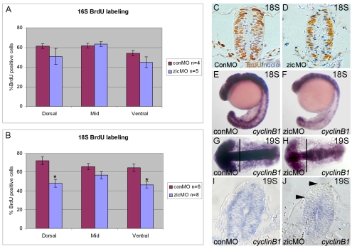
During neurulation, vertebrate embryos form a neural tube (NT), the rudiment of the central nervous system. In mammals and birds, a key step in cranial NT morphogenesis is dorsolateral hinge-point (DLHP) bending, which requires an apical actomyosin network. The mechanism of DLHP formation is poorly understood, although several essential genes have been identified, among them Zic2, which encodes a zinc-finger transcription factor. We found that DLHP formation in the zebrafish midbrain also requires actomyosin and Zic function. Given this conservation, we used the zebrafish to study how genes encoding Zic proteins regulate DLHP formation. We demonstrate that the ventral zic2a expression border predicts DLHP position. Using morpholino (MO) knockdown, we show zic2a and zic5 are required for apical F-actin and active myosin II localization and junction integrity. Furthermore, myosin II activity can function upstream of junction integrity during DLHP formation, and canonical Wnt signaling, an activator of zic gene transcription, is necessary for apical active myosin II localization, junction integrity and DLHP formation. We conclude that zic genes act downstream of Wnt signaling to control cytoskeletal organization, and possibly adhesion, during neurulation. This study identifies zic2a and zic5 as crucial players in the genetic network linking patterned gene expression to morphogenetic changes during neurulation, and strengthens the utility of the zebrafish midbrain as a NT morphogenesis model.


Figures

Fig. 1.
Fig. 1.
zic2a expression during zebrafish midbrain neurulation. (A) Schematic comparison between chick and zebrafish cranial neurulation. Presumptive dorsal tissue is shaded more darkly than ventral. DLHPs (asterisks) form before NT closure in chick, but after closure in zebrafish. (B-F) Wild-type zebrafish embryos stained at indicated stages for zic2a (purple) and for nuclei (red) by ISH with Neutral red. Ventral Zic expression border is labeled with black arrowheads. DLHPs form between 18-somite (18S) stage and 24 hpf. (G-J) Tg(zic2a-D5:egfp) embryos showing expression of GFP (green). (H,J) Phalloidin (red) counterstain. DLHPs (asterisks) form at the ventral border of GFP expression (white arrowheads). Images show midbrain cross-sections, dorsal at the top. Embryonic stages are listed in upper right corners.
Fig. 2.
Fig. 2.
zic2a, zic5 and actomyosin contraction are required for DLHP formation. Cross-sections through zebrafish midbrains stained with hematoxylin (purple) to label nuclei at 21-somite stage (A-E) and 24 hpf (F-J). (A) Control morphants show initial DLHP bending (asterisks). (B,C) The zic morphants have a small central lumen, but no DLHPs. (D) DMSO-treated controls display initial DLHP bending. (E) Blebbistatin-treated embryo with no DLHPs. (F) Control morphants have fully developed DLHPs. (G,H) The zic morphants show either an open lumen with no DLHPs (G) or a small lumen with very little DLHP bending (H). (I) DMSO controls form normal DLHPs. (J) Blebbistatin treatment inhibits DLHPs and prevents lumen opening.
Fig. 3.
Fig. 3.
zic2a and zic5 are required for apical F-actin localization and phosphorylated-myosin-II expression. (A-L) Horizontal sections through dorsal midbrain, anterior on the left, showing phalloidin (green) and rMLC-P (red) expression. At 16-somite stage, broad apical staining is seen in controls (A-C), but not zic morphants (D-F). By the 19-somite stage, a tight apical seam of staining is apparent in controls (G-I), but not in morphants (J-L). (M-R) Midbrain cross-sections, dorsal at the top, of Tg(zic2a-D5:egfp) embryos showing GFP (green) and phalloidin (red). (M-O) Controls show a contiguous apical seam of F-actin. (P-R) The zic morphants display disorganized phalloidin staining, particularly within the GFP domain. (C,I,O,F,L,R) Colored overlays of the images to the left. (S) 19-somite-stage zic morphants contain significantly less F-actin (*P<0.002) in the apical region than control morphants, based on measurements from high-magnification phalloidin-stained images. Error bars represent s.e.m. Representative images used for these measurements are shown in T (control morphant) and U (zic morphant). Scale bar: 20 μm. (V) Approximate location of the sections shown above. Arrowheads indicate the apical seam in C-L and dorsal apical seam in M and P.
Fig. 4.
Fig. 4.
Expression of apical junction markers in zic morphants. (A-L) Horizontal confocal sections through midbrain of immunolabeled embryos at 19-somite stage; anterior is on the left. (A-D) β-Catenin localizes to a contiguous apical domain in wild-type embryos (A,C). Apical β-catenin is patchy and discontinuous at the dorsal apical surface in zic morphants (B), but appears unaffected ventrally (D). Similar patterns are seen with Mpp5 (E-H) and ZO-1 (I-L). (M-N) Cross-sections through dorsal midbrain showing ZO-1 (red) and membrane-targeted mGFP (green) in controls (M) and zic morphants (N). (O) Approximate locations of the sections shown above.
Fig. 5.
Fig. 5.
Division orientation and GFP-Pk localization are normal in zic morphants. (A,B) Horizontal sections, anterior to the left, of 15-somite-stage embryos labeled with Histone2B:GFP (green) and phalloidin (red). Control (A) and zic morphant (B) embryos both show normal midline-crossing divisions. Lines drawn parallel to the midline and between anaphase chromosomes (white lines) were used to measure division orientation (inset in A). (C) Morphants and controls show comparable distributions of division angles, with the majority of cells dividing 80-120 degrees relative to the midline. (D-E) Horizontal sections, anterior to the left, of 12-somite-stage embryos scatter-labeled with GFP-Pk (green) and counterstained with phalloidin (red). Controls (D) and morphants (E) show cells with anteriorly biased GFP-Pk membrane puncta (arrowheads). (F) A comparison of GFP-Pk localization in controls and morphants.
Fig. 6.
Fig. 6.
The zic morphants have fewer S-phase cells and reduced ccnb1 transcript levels in the dorsal midbrain. (A) The percentage of BrdU-positive cells is not significantly reduced in zic morphants at 16-somite stage. (B) At 18-somite stage, zic morphants contain significantly fewer BrdU-positive cells in the dorsal midbrain (*P<0.002) and ventral midbrain (*P<0.008), determined by Student's t-test. To further investigate whether the number of S-phase cells was different across the dorsoventral axis for controls and zic morphants, we performed ANOVA analysis and found no significant difference (P=0.320). Error bars represent s.e.m. (C-D) Representative midbrain sections stained with anti-BrdU (brown) and hematoxylin (purple). (E-J) 18- to 19-somite-stage embryos stained by in situ hybridization for ccnb1. The zic morphants show a reduction in ccnb1 (F,H), primarily in the dorsal midbrain (between arrowheads in J). (E,F) Lateral views, anterior left. (G,H) Dorsal views, anterior left. (I,J) Representative cross-sections through midbrain.
Fig. 7.
Fig. 7.
Junction integrity, myosin activation and DLHP bending are normal in the absence of proliferation. (A-F) Immunostaining in horizontal confocal sections through dorsal midbrain at 20-somite stage. (A-B) Phosphohistone-H3 expression. C-D: ZO-1 expression. (E-F) rMLC-P expression. (G-H) The 24-hour midbrain cross-sections are labeled with phosphohistone-H3 (brown) and hematoxylin (purple). DMSO controls are shown on the left, aphidicolin-hydroxyurea treated embryos on the right. Asterisks indicate DLHPs. Arrowheads indicate the apical seam.
Fig. 8.
Fig. 8.
Cell proliferation and junction integrity require active actomyosin contraction. (A-F) Immunostaining in horizontal confocal sections through dorsal midbrain at 20-somite stage. (A-B) Phosphohistone-H3 expression. (C-D) ZO-1 expression. DMSO controls are shown on the left, blebbistatin on the right. Arrowheads indicate the apical seam.
Fig. 9.
Fig. 9.
Inhibition of Wnt signaling inhibits apical actomyosin contraction and disrupts apical junctions. (A-D) Immunostaining in horizontal confocal sections through dorsal midbrain at 21-somite stage. (A-B) rMLC-P expression. (C-D) ZO-1 expression. (E-F) 24 hpf midbrain cross-sections are labeled with hematoxylin (purple). All embryos were heat-shocked at 12-somite stage and recovered for 4 hours. Embryos carrying Tg(hs:gfpΔtcf) transgene are on the right, wild-type siblings on the left. Asterisks indicate DLHPs. Arrowheads mark the apical seam.
Fig. 10.
Fig. 10.
A molecular model of zic2a and zic5 in zebrafish DLHP development. Canonical Wnt signaling (blue) activates transcription of zic2a and zic5 (pink). The resultant Zic proteins regulate the apical actomyosin network, apical junction integrity and proliferation in the dorsal midbrain. Wnt signaling might also have zic-independent roles in these cellular processes (Aruga et al., 2002; Ebert et al., 2003). Actomyosin contraction and apical junction integrity cooperate to cause DLHP bending, forming a diamond-shaped lumen. Proliferation is not required for DLHP bending.

References

    1. Aaku-Saraste, E., Hellwig, A. and Huttner, W. B. (1996). Loss of occludin and functional tight junctions, but not ZO-1, during neural tube closure-remodeling of the neuroepithelium prior to neurogenesis. Dev. Biol. 180, 664-679. - PubMed
    1. Aruga, J. (2004). The role of Zic genes in neural development. Mol. Cell Neurosci. 26, 205-221. - PubMed
    1. Aruga, J., Nagai, T., Tokuyama, T., Hayashizaki, Y., Okazaki, Y., Chapman, V. M. and Mikoshiba, K. (1996). The mouse zic gene family: homologues of the Drosophila pair-rule gene odd-paired. J. Biol. Chem. 271, 1043-1047. - PubMed
    1. Aruga, J., Inoue, T., Hoshino, J. and Mikoshiba, K. (2002). Zic2 controls cerebellar development in cooperation with Zic1. J. Neurosci. 22, 218-225. - PMC - PubMed
    1. Bertet, C., Sulak, L. and Lecuit, T. (2004). Myosin-dependent junction remodelling controls planar cell intercalation and axis elongation. Nature 429, 667-671. - PubMed

Publication types

MeSH terms