. 2009 May;21(5):1541-55.
doi: 10.1105/tpc.108.064279. Epub 2009 May 26.

Remorin, a solanaceae protein resident in membrane rafts and plasmodesmata, impairs potato virus X movement

Affiliations

Remorin, a solanaceae protein resident in membrane rafts and plasmodesmata, impairs potato virus X movement

Sylvain Raffaele et al. Plant Cell. 2009 May.

Abstract

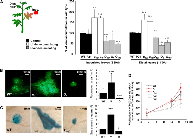
Remorins (REMs) are proteins of unknown function specific to vascular plants. We have used imaging and biochemical approaches and in situ labeling to demonstrate that REM clusters at plasmodesmata and in approximately 70-nm membrane domains, similar to lipid rafts, in the cytosolic leaflet of the plasma membrane. From a manipulation of REM levels in transgenic tomato (Solanum lycopersicum) plants, we show that Potato virus X (PVX) movement is inversely related to REM accumulation. We show that REM can interact physically with the movement protein TRIPLE GENE BLOCK PROTEIN1 from PVX. Based on the localization of REM and its impact on virus macromolecular trafficking, we discuss the potential for lipid rafts to act as functional components in plasmodesmata and the plasma membrane.


Figures

Figure 1.
Figure 1.
REM Is Located in the Cytosolic Leaflet of the PM and Is a Biochemical Marker of PM DIMs. (A) REM is found in the cytosolic leaflet of the PM. Representative protein gel blot (mean ± sd, n = 3) shows the detection of REM in phase partition-purified tobacco leaf PMs submitted to three subsequent cycles of freezing/thawing to progressively reverse the orientation of the RSO vesicles, followed by trypsin digestion. (B) Top: REM is a biochemical marker for plant DIMs. Isolation of DIMs from the PM using a TX100-to-protein ratio of 15. Ten fractions of equal volume were collected from the top to the bottom of the gradient. Precipitated proteins corresponding to half the volume of each fraction were analyzed by protein gel blots with antibodies to REM and to PMA. Bottom: Representative histogram shows total protein distribution along the gradient after DIM preparation (n = 10). (C) REM becomes TX100-soluble after mβCD treatment. PMs were treated or not with mβCD and submitted to the DIM isolation procedure. REM level was quantified in treated whole PMs (inset) and in the different fractions of the sucrose gradient by protein gel blots.
Figure 2.
Figure 2.
REM Locates in Membrane Domains in the Cytosolic Leaflet of the Tobacco Leaf PM. (A) Left: Transmission electron micrographs of negatively stained tobacco PM vesicles treated or not with mβCD (20 mM, 30 min). Center: REM locates in membrane domains of ∼80 nm. PM vesicles were treated or not with mβCD then immunogold labeled in batch, detected by GAR10, and observed by EM. Arrows point to obvious areas of REM clustering. Right: Statistical point patterns show analyses of the whole images by Ripley's K function. First minimum (arrowhead) of the derivative of K function (dotted) indicates the average size of clusters seen in the picture. (B) K-function analyses calculated as described in (A) are pooled and reported. (C) REM is clustered in DIMs on one side of the bilayer. PMs and DIMs processed as described in (A) were embedded in resin, ultrathin cut, and observed by EM. The PM appeared mainly as vesicles (V); DIMs appeared as membrane sheets more or less parallel to the section plane. Arrows show the border of the lipid raft bilayer.
Figure 3.
Figure 3.
Translational-GFP/YFP Fusions to REM Are Targeted to the PM. (A) Tobacco epidermis cells expressing the PM H+ pump ATPase (PMA4) fused to GFP (GFP-PMA4) and YFP-REM; observation by confocal microscopy shows that REM localizes to the PM. (B) Two-dimensional maximal projection of tobacco epidermis cell expressing GFP-REM showing labeling distribution through the whole cell; no labeling was observed in the cytoplasm (cytosol and organelles). (C) Detailed comparison between GFP-PMA and GFP-REM labeling in tobacco epidermis and BY-2 cells through PM and secant confocal planes. By contrast with GFP-PMA4, GFP-REM shows a patchy localization both in the PM plane and in close-up view through secant plane.
Figure 4.
Figure 4.
Translational-GFP/YFP Fusions to REM Localize Both in the PM and PD. (A) REM is located in adhesion sites (arrows) between the PM and cell wall in plasmolyzed calcofluor-stained (CaFl) BY-2 cells (left) or epidermal leaf tobacco cells (right) expressing GFP-REM. (B) REM colocalized with the PD marker PDLP1. Plasmolyzed tobacco cells expressing YFP-REM and GFP-PDLP1. Arrowheads show colabeled spots retained in the cell wall after plasmolysis, and asterisks indicate attachment sites to the retracted protoplast (see Supplemental Figure 5 online).
Figure 5.
Figure 5.
Endogenous REM Localizes Both in PM and PD in Vivo. (A) Localization of REM by immunofluorescence in tomato root cells. (B) Representative electron micrographs showing immunogold-labeled REM along the PM of high-pressure frozen tobacco root apices immunolabeled with antibodies to REMα130 and detected by GAR5 (arrowheads). (C) Representative electron micrographs and corresponding drawings showing immunogold-labeled REM in PD. Left: High-pressure frozen tobacco root apex immunolabeled with antibodies to REMα130 and detected by GAR5 (arrows). Right: Chemically fixed tomato shoot apex immunolabeled with antibodies to REMpp34 and detected by GAR10; arrowheads show the necks of PD. CW, cell wall.
Figure 6.
Figure 6.
Transgenic Tomato Lines with Altered REM Levels. (A) REM transgenes significantly impact REM accumulation in 1-month-old transgenic tomatoes. Lines overaccumulating (O) or underaccumulating REM (u) were selected. Wild-type and transgenic plants transformed with the empty vector (P21) were also analyzed. Protein gel blot on crude total protein extract from T2 transgenic tomato leaves (further used in Figure 7 experiments; at least six plants per line) was quantified and expressed as percentage of signal in the wild type; error bars show sd. Inset: The PM was purified from some T2 transgenic tomato leaves, and REM level was measured by protein gel blotting (B) Modulation of REM level in tomato causes no significant alteration in plant development except a slight early senescence in overaccumulating plants.
Figure 7.
Figure 7.
REM Misexpression Alters PVX Virus Propagation. (A) PVX viral charge is inversely correlated with REM level in 4-week-old transgenic tomatoes, both at the local and systemic levels. Viral charge was assayed by ELISA using antibodies to PVX coat protein on inoculated leaves (at 8 DAI) and distal (n+3) leaves (14 DAI). Three independent experiments were performed with seven to eight plants for each transgenic line and nontransgenic (WT) or empty vector control (P21). (B) and (C) PVX spreading was inversely correlated with REM level in transgenic tomatoes. Four-week-old wild-type and transgenic tomato lines were inoculated with PVX-GFP (B) or PVX-GUS (C). Representative pictures of inoculated leaves were taken at 4 DAI. Graphs show average area of infection foci from ∼150 pictures of various lines of each genotype. (D) Protoplasts of transgenic tomatoes misexpressing REM were inoculated with 4 μg of purified PVX RNA. PVX RNA accumulation was measured by qRT-PCR and normalized to 25S rRNA. HAI, hours postinoculation. For graphs, error bars show sd, and significance is assessed by a Student's t test (*, P < 0.1; **, P < 0.05; ***, P < 0.001).
Figure 8.
Figure 8.
REM Directly Binds PVX TGBp1 to Alter PVX Cell-to-Cell Transfer. (A) REM directly interacts with TGBp1 in a yeast split ubiquitin assay. Split ubiquitin interaction between REM and PVX movement (TGBP-1, -2, and -3) and capsid (CP) proteins. (B) REM directly interacts with TGBp1 in a pull-down assay. GFP-tagged TGP1-2 and CP were transiently expressed in tobacco leaves. Leaf crude extracts were analyzed by protein gel blots using antibodies to GFP. Proteins were extracted and incubated overnight with glutathione sepharose beads with or without GST-tagged REM. Pulled-down proteins were analyzed after three washes. (C) YFP-REM and TGBp1-GFP colocalize when transiently expressed in tobacco leaves. Image shows two-dimensional maximal projections throughout the cell.

Comment in

References

    1. Bariola, P.A., Retelska, D., Stasiak, A., Kammerer, R.A., Fleming, A., Hijri, M., Frank, S., and Farmer, E.E. (2004). Remorins form a novel family of coiled coil-forming oligomeric and filamentous proteins associated with apical, vascular and embryonic tissues in plants. Plant Mol. Biol. 55 579–594. - PubMed
    1. Batoko, H., Zheng, H.Q., Hawes, C., and Moore, I. (2000). A rab1 GTPase is required for transport between the endoplasmic reticulum and golgi apparatus and for normal golgi movement in plants. Plant Cell 12 2201–2218. - PMC - PubMed
    1. Benitez-Alfonso, Y., Cilia, M., San Roman, A., Thomas, C., Maule, A., Hearn, S., and Jackson, D. (2009). Control of Arabidopsis meristem development by thioredoxin-dependent regulation of intercellular transport. Proc. Natl. Acad. Sci. USA 106 3615–3620. - PMC - PubMed
    1. Bhat, R.A., Miklis, M., Schmelzer, E., Schulze-Lefert, P., and Panstruga, R. (2005). Recruitment and interaction dynamics of plant penetration resistance components in a plasma membrane microdomain. Proc. Natl. Acad. Sci. USA 102 3135–3140. - PMC - PubMed
    1. Bhat, R.A., and Panstruga, R. (2005). Lipid rafts in plants. Planta 223 5–19. - PubMed

Publication types

MeSH terms