

Structural specializations of A2, a force-sensing domain in the ultralarge vascular protein von Willebrand factor
- PMID: 19470641
- PMCID: PMC2695068
- DOI: 10.1073/pnas.0903679106
Structural specializations of A2, a force-sensing domain in the ultralarge vascular protein von Willebrand factor
Abstract
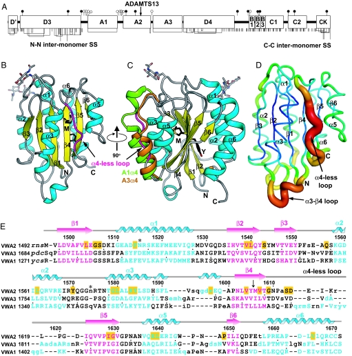
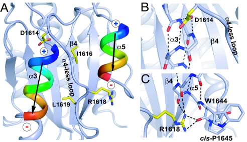
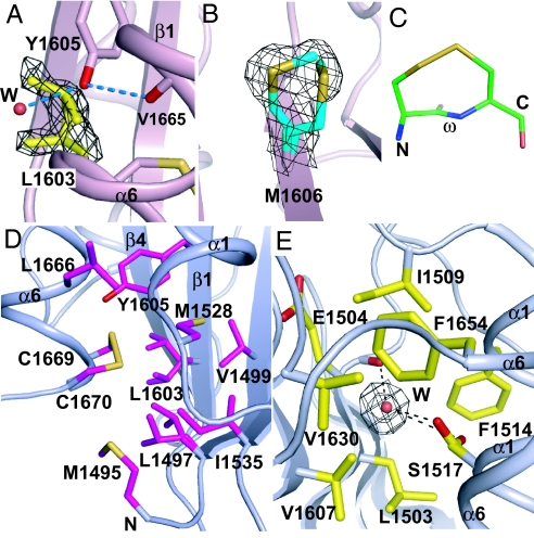
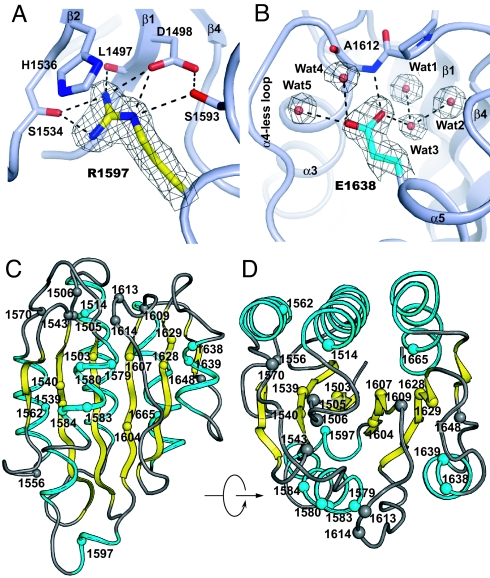
The lengths of von Willebrand factor (VWF) concatamers correlate with hemostatic potency. After secretion in plasma, length is regulated by hydrodynamic shear force-dependent unfolding of the A2 domain, which is then cleaved by a specific protease. The 1.9-A crystal structure of the A2 domain demonstrates evolutionary adaptations to this shear sensor function. Unique among VWF A (VWA) domains, A2 contains a loop in place of the alpha4 helix, and a cis-proline. The central beta4-strand is poorly packed, with multiple side-chain rotamers. The Tyr-Met cleavage site is buried in the beta4-strand in the central hydrophobic core, and the Tyr structurally links to the C-terminal alpha6-helix. The alpha6-helix ends in 2 Cys residues that are linked by an unusual vicinal disulfide bond that is buried in a hydrophobic pocket. These features may narrow the force range over which unfolding occurs and may also slow refolding. Von Willebrand disease mutations, which presumably lower the force at which A2 unfolds, are illuminated by the structure.
Conflict of interest statement
The authors declare no conflict of interest.
Figures
References
-
- Mayans O, et al. Structural basis for activation of the titin kinase domain during myofibrillogenesis. Nature. 1998;395:863–869. - PubMed
-
- Sadler JE. Biochemistry and genetics of von Willebrand factor. Annu Rev Biochem. 1998;67:395–424. - PubMed
-
- Ruggeri ZM, Mendolicchio GL. Adhesion mechanisms in platelet function. Circ Res. 2007;100:1673–1685. - PubMed
-
- Batlle J, et al. Proteolytic degradation of von Willebrand factor after DDAVP administration in normal individuals. Blood. 1987;70:173–176. - PubMed
-
- Dong JF, et al. ADAMTS-13 rapidly cleaves newly secreted ultralarge von Willebrand factor multimers on the endothelial surface under flowing conditions. Blood. 2002;100:4033–4039. - PubMed
Publication types
MeSH terms
Substances
Associated data
- Actions
Grants and funding
LinkOut - more resources
Full Text Sources
Other Literature Sources
Miscellaneous
