. 2009 Jun 9;106(23):9215-20.
doi: 10.1073/pnas.0811070106. Epub 2009 May 21.

Alteration of enzyme specificity by computational loop remodeling and design

Affiliations

Alteration of enzyme specificity by computational loop remodeling and design

Paul M Murphy et al. Proc Natl Acad Sci U S A. .

Abstract

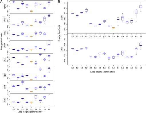
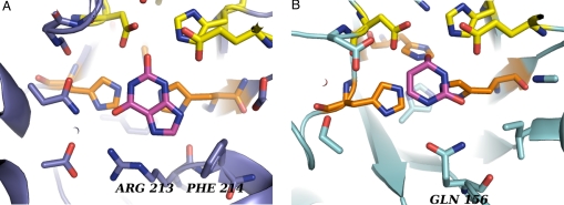
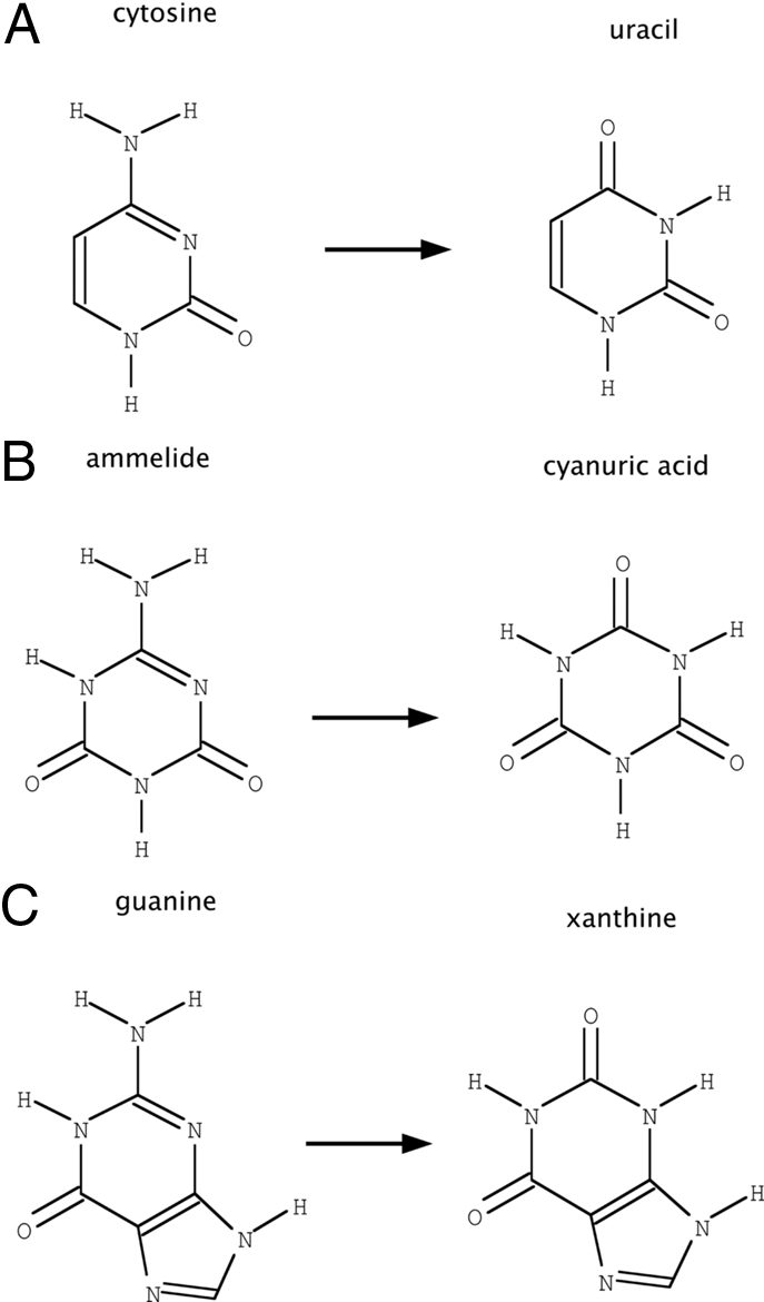
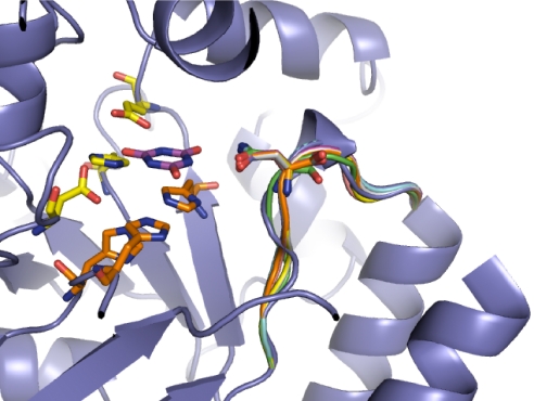
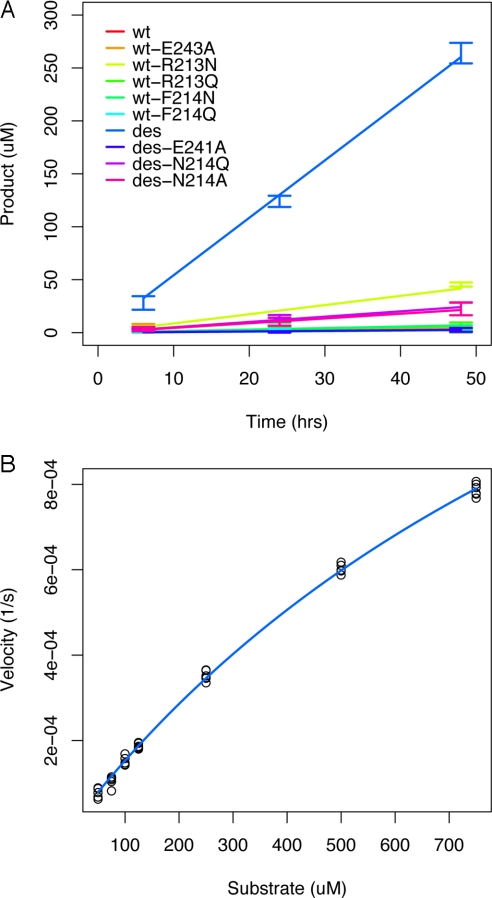
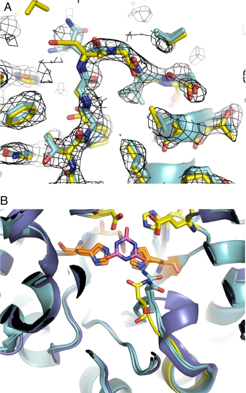
Altering the specificity of an enzyme requires precise positioning of side-chain functional groups that interact with the modified groups of the new substrate. This requires not only sequence changes that introduce the new functional groups but also sequence changes that remodel the structure of the protein backbone so that the functional groups are properly positioned. We describe a computational design method for introducing specific enzyme-substrate interactions by directed remodeling of loops near the active site. Benchmark tests on 8 native protein-ligand complexes show that the method can recover native loop lengths and, often, native loop conformations. We then use the method to redesign a critical loop in human guanine deaminase such that a key side-chain interaction is made with the substrate ammelide. The redesigned enzyme is 100-fold more active on ammelide and 2.5e4-fold less active on guanine than wild-type enzyme: The net change in specificity is 2.5e6-fold. The structure of the designed protein was confirmed by X-ray crystallographic analysis: The remodeled loop adopts a conformation that is within 1-A Calpha RMSD of the computational model.


Conflict of interest statement

The authors declare no conflict of interest.

Figures

Fig. 1.
Fig. 1.
Benchmarking and application of loop design methodology. (A) Eight protein/ligand complexes were chosen from the PDB to benchmark the loop design protocol. A 7-residue window centered on an anchor residue was excised from a model of the complex. To assess our ability to recapitulate native loop lengths with this protocol, the excised region was filled by using loops of the native length (3,3; orange) and several nonnative lengths (blue). Loop lengths indicate the number of residues inserted in the excised region before and after the anchor residue. The total loop length also includes the anchor residue. The energies of the 5 lowest-energy structures generated for each loop length are shown in box-and-whiskers form. In 6 of 8 cases, the native length structures are lower in energy than the nonnative length structures. Thus, native loop lengths can be identified with this protocol. (B) The sequence requirements for hGDA specificity alteration were determined by comparing energies of several different loop lengths and anchor identities. The lowest-energy structures (orange) had loop length 3,4, which was 2 residues shorter than the native loop, and anchor identity asparagine. The lowest-energy glutamine design had loop length 2,4 and had a higher energy than the lowest-energy asparagine design. The lowest energy loop overall (orange, asparagine) was selected for experimental characterization.
Fig. 2.
Fig. 2.
Active-site structures. (A) The active site of human guanine deaminase (hGDA) with product, xanthine (PDB ID code 2UZ9). Arginine 213 and phenylalanine 214 are visible at the bottom of the image. (B) The active site of bacterial cytosine deaminase (bCD) with transition state analog, di-hydropyrimidine (PDB ID code 1K70). Glutamine 156 is visible at the bottom of the image. Proton shuttling residues, colored yellow, are conserved and are responsible for positioning and transferring protons from a bound water molecule to the substrate. Metal-binding histidines, colored orange, are responsible for binding a metal (zinc in hGDA, iron in bCD), which lowers the pKa of the bound water molecule. The transition state of the reaction is the tetrahedral intermediate formed after attack of the bound water molecule and before leaving group departure.
Fig. 3.
Fig. 3.
Deaminase substrates. (A) The reaction performed by wild-type human guanine deaminase (hGDA). (B) The reaction under study in this article. (C) The reaction performed by prodrug-activating cytosine deaminases, such as bacterial cytosine deaminase (bCD). Ammelide is a structural intermediate between guanine and cytosine and thus provides a stepping stone for specificity alteration of hGDA. Each reaction consumes 1 molecule of H2O and releases 1 molecule of NH3.
Fig. 4.
Fig. 4.
Structure of design models. The backbone configuration of several designs are superimposed on wild-type hGDA, highlighting the differences that allow binding to the new reaction transition state. The design structures differ in length, conformation, and sequence. Two residues have been deleted, and 4 mutations have been made, including the introduction of an asparagine that directly interacts with the substrate.
Fig. 5.
Fig. 5.
Kinetic characterization. (A) Product formation over time at 20 μM enzyme, 500 μM substrate reveals that hGDA-des is highly active relative to wild type (100-fold), and asparagine or glutamine mutants without the remodeled backbone (>6-fold, Table S3). Mutations of the designed asparagine to alanine or glutamine demonstrates significantly reduced activity. (B) Velocity vs. substrate concentration at enzyme concentration of 10 μM. The estimated Michaelis–Menten parameters of hGDA-des are: kcat = 2.2 (2.1–2.4) e−4 s−1, Km = 1,300 (1,200–1,500) μM. Estimating kcat/Km from the 4 lowest substrate concentrations gives 0.15 (0.14–0.15) s−1 M−1 (95% confidence intervals in parentheses).
Fig. 6.
Fig. 6.
Superimposition of computational model and crystal structure. In both A and B, the computational model of hGDA-des is shown in yellow, whereas both chains of the final refined structure are shown in cyan. The backbone of the computational model is within 1-Å Cα RMSD of the crystal structure. (A) Unbiased electron density using phases derived from molecular replacement is shown at the 1σ level in mesh. The backbone of the designed loop is clearly visible in the density after molecular replacement. The side chain of designed residue N214 was not clearly visible in the electron density at this stage or after refinement. The side chain of designed residue V216 is clearly visible in the expected location. (B) No electron density was observed in the location expected for bound substrate, ammelide, or product, cyanuric acid, but for comparison, the modeled location is shown in magenta. The wild-type structure is also shown in slate blue.

References

    1. Dahiyat BI, Mayo SL. De novo protein design: Fully automated sequence selection. Science. 1997;278:82–87. - PubMed
    1. Kuhlman B, et al. Design of a novel globular protein fold with atomic-level accuracy. Science. 2003;302:1364–1368. - PubMed
    1. Looger LL, et al. Computational design of receptor and sensor proteins with novel functions. Nature. 2003;423:185–190. - PubMed
    1. Kortemme T, et al. Computational redesign of protein–protein interaction specificity. Nat Struct Mol Biol. 2004;11:371–379. - PubMed
    1. Ashworth J, et al. Computational redesign of endonuclease DNA binding and cleavage specificity. Nature. 2006;441:656–659. - PMC - PubMed

Publication types

Associated data