. 2009 Sep;19(9):971-86.
doi: 10.1093/glycob/cwp074. Epub 2009 May 21.

Mutational and functional analysis of Large in a novel CHO glycosylation mutant

Affiliations

Mutational and functional analysis of Large in a novel CHO glycosylation mutant

Jennifer T Aguilan et al. Glycobiology. 2009 Sep.

Abstract

Inactivating mutations of Large reduce the functional glycosylation of alpha-dystroglycan (alpha-DG) and lead to muscular dystrophy in mouse and humans. The N-terminal domain of Large is most similar to UDP-glucose glucosyltransferases (UGGT), and the C-terminal domain is related to the human i blood group transferase beta1,3GlcNAcT-1. The amino acids at conserved motifs DQD+1 and DQD+3 in the UGGT domain are necessary for mammalian UGGT activity. When the corresponding residues were mutated to Ala in mouse Large, alpha-DG was not functionally glycosylated. A similar result was obtained when a DXD motif in the beta1,3GlcNAcT-1 domain was mutated to AIA. Therefore, the first putative glycosyltransferase domain of Large has properties of a UGGT and the second of a typical glycosyltransferase. Co-transfection of Large mutants affected in the different glycosyltransferase domains did not lead to complementation. While Large mutants were more localized to the endoplasmic reticulum than wild-type Large or revertants, all mutants were in the Golgi, and only very low levels of Golgi-localized Large were necessary to generate functional alpha-DG. When Large was overexpressed in ldlD.Lec1 mutant Chinese hamster ovary (CHO) cells which synthesize few, if any, mucin O-GalNAc glycans and no complex N-glycans, functional alpha-DG was produced, presumably by modifying O-mannose glycans. To investigate mucin O-GalNAc glycans as substrates of Large, a new CHO mutant Lec15.Lec1 that lacked O-mannose and complex N-glycans was isolated and characterized. Following transfection with Large, Lec15.Lec1 cells also generated functionally glycosylated alpha-DG. Thus, Large may act on the O-mannose, complex N-glycans and mucin O-GalNAc glycans of alpha-DG.


Figures
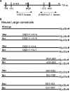
Fig. 1
Fig. 1
Diagram of Large and mutant constructs. (A) Mouse Large is 756 amino acids including a transmembrane domain (TM, aa 12-34), coiled coil domain 1 (CC1, aa 55–90), coiled coil domain 2 (CC2, aa 483–496), and five putative N-glycosylation sites (*). There are four DXD motifs at aa 242–244 (DTD, not shown), aa 334–336 (DQD), aa 438–440 (DED, not shown), and aa 563–565 (DID). The DQD, DQD+1(I), and DQD+3(N) residues are conserved in the C-terminal domain of UGGTs. The DTD and DQD are also spaced similarly in Large and UGGTs. The DID at aa 563–565 is conserved in mouse and human β3GlcNAcT-1 (or iGnT1), which initiates extension of polylactosamine units. (B) Constructs of Large and Large mutants with MycHis tag (⧫) in the pIRES-GFP vector.
Fig. 2
Fig. 2
Mutations that inactivate Large. (A) Lec8 cells were transiently transfected with the pIRES-eGFP vector (GFP) or wild-type (WT) Large, Large mutants, or revertants (-R). Lysates were subjected to Western analysis, and blots were cut and probed separately with mAb IIH6, or anti-Myc mAb, or anti-β-DG. The laminin overlay assay was performed on the same amount of the same cell lysates. (B) Co-transfection of two Large constructs together was performed as shown and lysates analyzed as in (A).
Fig. 3
Fig. 3
Subcellular localization of Large and Large mutants. (A) Wild-type Large and Large mutants with C-terminal Myc were detected using anti-Myc mAb and secondary antibody conjugated to AlexaFluor 568 (red). DAPI (blue) was used for nuclear staining. The TGN38 Golgi marker and PDI ER markers were detected using secondary Ab conjugated to AlexaFluor488 (green). First row: localization of WT Large and TGN38. Second row: localization of WT Large and PDI. Third row: Large showing enrichment in both the Golgi and ER compartments. Fourth row: Large AIA mutant with reticular-like staining merged with PDI. (B) Percent transfectants expressing wild-type Large or Large mutants and revertants in Golgi, Golgi and ER (Golgi/ER) or ER (n ≥ 100 for each).
Fig. 4
Fig. 4
Titration of Large cDNA. (A) Decreasing amounts of Large cDNA supplemented to a total of 4 μg cDNA with pIRES-eGFP cDNA were transfected into CHO, and lysates were analyzed by Western analysis using mAb IIH6, stripped, and re-probed with anti-Myc mAb. The laminin overlay assay was performed on a second portion of the same cell lysates. (B) The same experiment was performed with Lec8 cells.
Fig. 5
Fig. 5
Effects of Large on α-DG in ldlD.Lec1 cells. (A) VVL binding to blots of cell lysates of ldlD.Lec1 grown in alpha-MEM with 10% dialyzed fetal bovine serum without exogenous sugars for 4 days and Lec8 and CHO grown in alpha-MEM with 10% fetal bovine serum. (B) DSA lectin binding to cell lysates from CHO, Lec8, and ldlD.Lec1 grown as in (A). (C) PNA binding to cell lysates from Lec2 and ldlD.Lec1 grown in serum-free medium (30 min exposure). (D) Western analysis of cell lysates from ldlD.Lec1 and Lec2 cells transfected with Large or pIRES-GFP vector and grown in serum-free medium. Blots were cut and probed separately with mAbs IIH6, antiMyc, and anti-β-DG. The laminin overlay assay was performed using another portion of the same cell lysates.
Fig. 6
Fig. 6
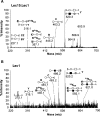
MS/MS analysis of precursor ion m/z 738 from Lec15.Lec1 and Lec1. (A) MALDI-TOF/TOF analysis of the precursor ion m/z 738 from the MALDI-TOF spectrum of Lec15.Lec1 permethylated O-glycans (see supplementary Figure S1) with fragment ions assigned using Glycoworkbench. (B) MALDI-TOF/TOF analysis of the precursor ion m/z 738 from the MALDI-TOF spectrum of Lec1 permethylated O-glycans (see supplementary Figure S1). Fragment ions show a mixture of isobaric structures corresponding to Hex2HexNAc-ol (e.g., Gal-Gal-GalNAc-ol) mucin O-glycans and HexHexNAcHex-ol (e.g., Gal-GlcNAc-Man-ol). Sialic acid (◊); Gal (formula image); GlcNAc (▪); Man (formula image).
Fig. 7
Fig. 7
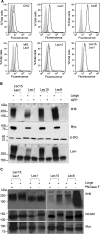
Cell surface expression of endogenous α-DG and effects of Large on α-DG in Lec15.Lec1 cells. (A) Flow cytometry using primary antibody 6C1 anti-cranin (α-DG) and secondary antibody FITC-conjugated to anti-mouse IgG+A+M (black line), secondary Ab alone (gray profile), and primary antibody alone (dark gray line). (B) Cell lysates of Lec8, Lec15, Lec1, and Lec15.Lec1 cells expressing Large or pIRES-GFP vector control and probed using primary mAb IIH6, anti-Myc, or anti-β-DG followed by HRP-conjugated goat anti-mouse IgG+M Ab. The laminin overlay assay was performed using another portion of the same cell lysates. (C) Another portion of the same cell lysates was analyzed before and after treatment with PNGase F. PNGase F digestion was confirmed by stripping the membrane and probing with anti-NCAM, and expression levels of Large by stripping the same membrane again and probing with anti-Myc mAb.
Fig. 8
Fig. 8
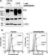
Effects of AcBenzyl-O-GalNAc on the functional glycosylation of α-DG by Large. (A) Lec8 and Lec15.Lec1 cells were incubated for 24 h in the presence or absence of 1 mM AcBzOGalNAc and transfected with Large for the next 24 h. Western analysis of lysates was performed using mAbs IIH6, anti-Myc, and anti-β-DG. (B) Lec8 and Lec15.Lec1 cells were incubated 24 h in the presence and absence of 1 mM AcBzOGalNAc, transfected with Large and incubated another 24 h before preparation for flow cytometry. The profiles show binding to primary antibody 6C1 mAb and secondary antibody FITC-conjugated to anti-mouse IgG+A+M. Secondary Ab alone (gray).
Fig. 9
Fig. 9
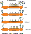
Large modifies different mammalian glycans. The diagram shows a model of α-DG with the range of glycans it is proposed to carry when synthesized in CHO cells or the mutants Lec8, ldlD.Lec1, or Lec15.Lec1. Large generates functionally glycosylated α-DG when produced in each of these cell lines. In CHO and Lec8 cells, Large-modified N-glycans are sensitive to treatment with PNGase F. Large-modified N-glycans are not sensitive to PNGase F in ldlD.Lec1 or Lec15.Lec1 and thus Large modifies complex N-glycans but not oligomannosyl N-glycans. In ldlD.Lec1 cells, the major substrate of Large is O-mannose glycans because O-GalNAc glycans are not synthesized and oligomannosyl N-glycans are not a substrate. In Lec15.Lec1 cells, the major substrate of Large is O-GalNAc glycans because O-mannose glycans are not synthesized, and oligomannosyl N-glycans are not a substrate. We propose that Large transfers either a Glc, GlcNAc, or GlcUA in α1,3-linkage to the hydroxyl at C6 of GlcNAc or GalNAc. The C6 hydroxyl is proposed because Large modifies glycans in which GlcNAc or GalNAc is substituted at the C2, C3, and/or C4 positions.

References

    1. Aoki K, Porterfield M, Lee SS, Dong B, Nguyen K, McGlamry KH, Tiemeyer M. The diversity of O-linked glycans expressed during Drosophila melanogaster development reflects stage- and tissue-specific requirements for cell signaling. J Biol Chem. 2008;283:30385–30400. - PMC - PubMed
    1. Arnold SM, Fessler LI, Fessler JH, Kaufman RJ. Two homologues encoding human UDP-glucose:glycoprotein glucosyltransferase differ in mRNA expression and enzymatic activity. Biochemistry. 2000;39:2149–2163. - PubMed
    1. Arnold SM, Kaufman RJ. The noncatalytic portion of human UDP-glucose: Glycoprotein glucosyltransferase I confers UDP-glucose binding and transferase function to the catalytic domain. J Biol Chem. 2003;278:43320–43328. - PubMed
    1. Barresi R, Campbell KP. Dystroglycan: From biosynthesis to pathogenesis of human disease. J Cell Sci. 2006;119:199–207. - PubMed
    1. Barresi R, Michele DE, Kanagawa M, Harper HA, Dovico SA, Satz JS, Moore SA, Zhang W, Schachter H, Dumanski JP, et al. LARGE can functionally bypass alpha-dystroglycan glycosylation defects in distinct congenital muscular dystrophies. Nat Med. 2004;10:696–703. - PubMed

Publication types