. 2009 Jul;19(7):1279-88.
doi: 10.1101/gr.089417.108. Epub 2009 May 21.

Targeted genome editing in human cells with zinc finger nucleases constructed via modular assembly

Affiliations

Targeted genome editing in human cells with zinc finger nucleases constructed via modular assembly

Hye Joo Kim et al. Genome Res. 2009 Jul.

Abstract

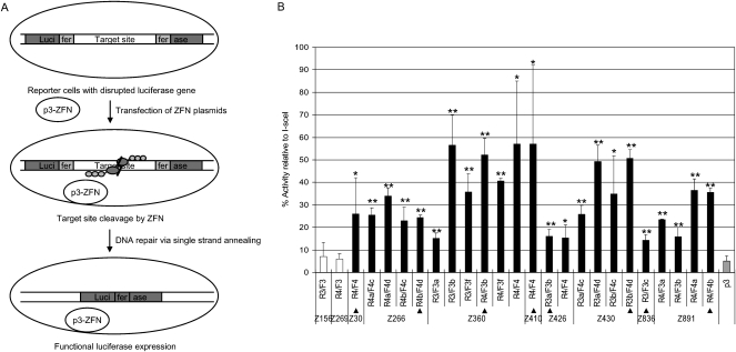
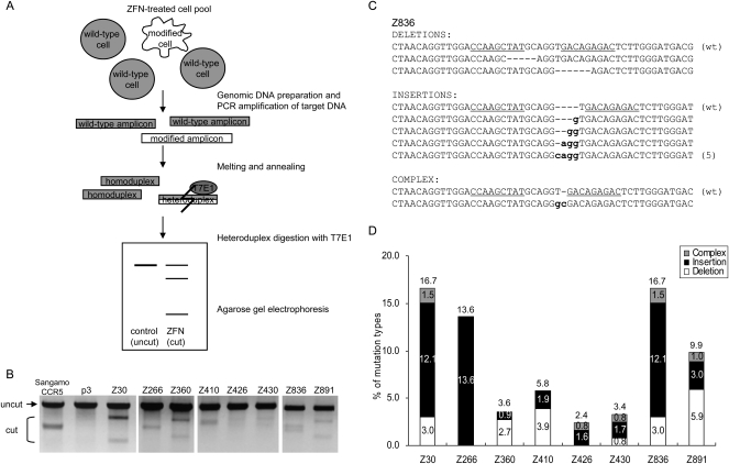
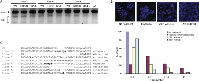
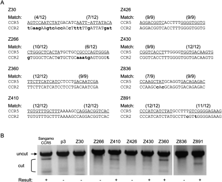
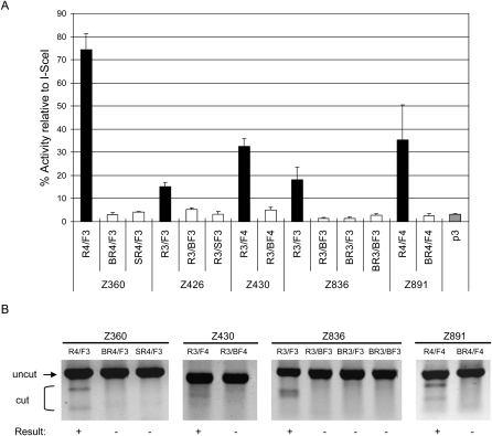
Broad applications of zinc finger nuclease (ZFN) technology-which allows targeted genome editing-in research, medicine, and biotechnology are hampered by the lack of a convenient, rapid, and publicly available method for the synthesis of functional ZFNs. Here we describe an efficient and easy-to-practice modular-assembly method using publicly available zinc fingers to make ZFNs that can modify the DNA sequences of predetermined genomic sites in human cells. We synthesized and tested hundreds of ZFNs to target dozens of different sites in the human CCR5 gene-a co-receptor required for HIV infection-and found that many of these nucleases induced site-specific mutations in the CCR5 sequence. Because human cells that harbor CCR5 null mutations are functional and normal, these ZFNs might be used for (1) knocking out CCR5 to produce T-cells that are resistant to HIV infection in AIDS patients or (2) inserting therapeutic genes at "safe sites" in gene therapy applications.


Figures

Figure 1.
Figure 1.
Assessment of ZFN activities using a cell-based single-strand annealing (SSA) system. (A) Schematic overview of a single-strand annealing system. ZFN expression plasmids are transfected into HEK293 cells whose genome contains an inactive, partially duplicated, and disrupted luciferase gene. If ZFNs cleave target sites in cells, DNA is efficiently repaired by the SSA mechanism, and the functional luciferase gene is restored. (B) Luciferase activities of cells in which various ZFNs are expressed. p3 (gray bar) is the empty plasmid, which was used as a negative control. The target sequence contains the recognition site of I-SceI, which was used as a positive control. The activity of each ZFN pair is reported as the percentage relative to the I-SceI control. ZFN pairs and their constituent monomers are indicated. The ZFN pairs used in further studies are marked with triangles. Means and standard deviations (error bars) from at least three independent experiments are shown. Black and white bars indicate active and inactive ZFNs, respectively. P-values were calculated with the Student's t-test; (*) P < 0.01; (**) P < 0.001 (p3 control vs. ZFN).
Figure 2.
Figure 2.
ZFN-mediated genome editing in human cells. (A) Schematic overview of mismatch detection using T7E1 assay. Genomic DNA was purified from cells transfected with plasmids encoding ZFNs. The DNA segments encompassing the sites of ZFN recognition were PCR-amplified, and the DNA amplicons were melted and annealed. If the DNA amplicons contain both wild-type and mutated DNA sequences, heteroduplexes would be formed. T7E1 recognizes and cleaves heteroduplexes, but not homoduplexes. The DNA fragments were assessed by agarose gel electrophoresis; a schematic of an idealized gel result is shown. (B) ZFN-mediated genomic modification revealed by the T7E1 assay. The ZFN pairs are shown at the top of the agarose gels. The expected positions of the resulting DNA bands are indicated by an arrow (uncut) and a bracket (cut) at the left of the gel panels. p3 is the empty plasmid used as a negative control. Sangamo's CCR5-targeting ZFN pair (Sangamo CCR5) was included in this assay. (C) DNA sequences of a genomic site targeted by the Z836 ZFN pair. (Underlined) ZFN recognition elements. (Dashes) Deletions; (small letters) inserted bases. In cases in which a mutation was detected more than once, the number of occurrences is shown in parentheses. (wt) Wild type. (D) Types of mutations at various ZFN-targeted sites. The number of deletions, insertions, and complex mutations for each ZFN pair (as shown in C for Z836) were counted, and the percentages of these incidents were plotted.
Figure 3.
Figure 3.
Obligatory heterodimeric ZFNs and isolation of mutant clones. (A) Time-course analysis of wild-type and obligatory heterodimeric ZFNs. The T7E1 mismatch detection assay was performed at various time points with DNA isolated from cells treated with different forms of the FokI nuclease domain. (WT) Wild-type nuclease domain; (RR/DD or KK/EL) obligatory heterodimeric nuclease domains. (B) ZFN-induced DSBs detected by TP53BP1 staining. Both the wild-type and obligatory heterodimeric Z891 ZFN pair were transfected into HEK293T/17 cells, and intracellular TP53BP1 foci were detected by immunofluorescence at day 2 post-transfection. Etoposide (1 μM) was used as a positive control. The distribution of the numbers of TP53BP1 foci is plotted. At least 100 cells were analyzed for each treatment in two independent measurements. (C) DNA sequences of mutant clones. Seven mutant clones were isolated, by limiting dilution, from cells treated with the RR/DD ZFN dimer. The DNA sequences of the target site in these clonal cells are shown. (Dashes) Deletions are indicated with dashes; (small letters) inserted bases. Clones 1a and 1b indicate DNA sequences that resulted from biallelic modifications in a single clone.
Figure 4.
Figure 4.
Off-target effects of ZFNs at the CCR2 locus. (A) ZFN recognition elements at the CCR5 and CCR2 loci. The ZFN pairs are indicated at the left of the DNA sequences. (Parentheses) The numbers of base matches between the CCR5 and CCR2 loci; (lowercase bold letters) mismatched bases; (underlined) the half-site ZFN recognition elements. (B) T7E1 assay at CCR2 sites. PCR-amplified DNA corresponding to the CCR2 coding region from cells treated with the ZFN pairs (shown at the top of the gel panels) was analyzed. (+) ZFN pairs that gave rise to the modification at the CCR2 sites. p3 is the empty plasmid used as a negative control. Sangamo's CCR5-targeting ZFN pair (Sangamo CCR5) was included in this assay.
Figure 5.
Figure 5.
Module swap analysis. New ZFNs were prepared using engineered ZFs to test whether these ZFNs can functionally replace ZFNs composed exclusively of naturally occurring modules that recognize identical sequences. The names of these ZFNs are indicated by the Z numbers at (A) the bottom of the graph or (B) the top of the gel panels. The ZFN monomers whose names start with “B,” such as “BR4,” are composed exclusively of Barbas modules, and those with “S,” such as “SR4,” are composed exclusively of Sangamo modules. The ZFs that compose these ZFNs are described in Supplemental Table 4. These ZFNs were analyzed using (A) the cell-based SSA system and (B) the T7E1 assay. (A) Luciferase activities of cells in which the various ZFNs were expressed. (Gray bar) p3 is the empty plasmid, which was used as a negative control. The target sequence contains the recognition site of I-SceI, which was used as a positive control. The activity of each ZFN pair is reported as the percentage relative to the I-SceI control. ZFN pairs and their constituent monomers are indicated. Means and standard deviations (error bars) from at least three independent experiments are shown. (B) ZFN-mediated genomic modification revealed by the T7E1 assay. ZFN pairs and their constituent monomers are shown at the top of the agarose gels. (+) ZFN pairs that gave rise to positive gene-editing events.

References

    1. Agrawal L, Lu X, Qingwen J, VanHorn-Ali Z, Nicolescu IV, McDermott DH, Murphy PM, Alkhatib G. Role for CCR5Δ32 protein in resistance to R5, R5X4, and X4 human immunodeficiency virus type 1 in primary CD4+ cells. J Virol. 2004;78:2277–2287. - PMC - PubMed
    1. Bae KH, Kim JS. One-step selection of artificial transcription factors using an in vivo screening system. Mol Cells. 2006;21:376–380. - PubMed
    1. Bae KH, Kwon YD, Shin HC, Hwang MS, Ryu EH, Park KS, Yang HY, Lee DK, Lee Y, Park J, et al. Human zinc fingers as building blocks in the construction of artificial transcription factors. Nat Biotechnol. 2003;21:275–280. - PubMed
    1. Beerli RR, Segal DJ, Dreier B, Barbas CF., III Toward controlling gene expression at will: Specific regulation of the erbB-2/HER-2 promoter by using polydactyl zinc finger proteins constructed from modular building blocks. Proc Natl Acad Sci. 1998;95:14628–14633. - PMC - PubMed
    1. Beumer K, Bhattacharyya G, Bibikova M, Trautman JK, Carroll D. Efficient gene targeting in Drosophila with zinc-finger nucleases. Genetics. 2006;172:2391–2403. - PMC - PubMed

LinkOut - more resources