. 2009 Jun 1;15(11):3740-50.
doi: 10.1158/1078-0432.CCR-08-3252. Epub 2009 May 26.

HGF and c-Met participate in paracrine tumorigenic pathways in head and neck squamous cell cancer

Affiliations

HGF and c-Met participate in paracrine tumorigenic pathways in head and neck squamous cell cancer

Lynn M Knowles et al. Clin Cancer Res. .

Abstract

Purpose: We determined hepatocyte growth factor (HGF) and c-Met expression and signaling in human head and neck squamous cell carcinoma (HNSCC) cells and primary tissues and tested the ability of c-Met tyrosine kinase inhibitors (TKI) to block HGF-induced biological signaling.

Experimental design: Expression and signaling were determined using immunoblotting, ELISA, and immunohistochemistry. Biological end points included wound healing, cell proliferation, and invasion. c-Met TKIs were tested for their ability to block HGF-induced signaling and biological effects in vitro and in xenografts established in nude mice.

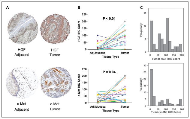
Results: c-Met was expressed and functional in HNSCC cells. HGF was secreted by HNSCC tumor-derived fibroblasts, but not by HNSCC cells. Activation of c-Met promoted phosphorylation of AKT and mitogen-activated protein kinase as well as release of the inflammatory cytokine interleukin-8. Cell growth and wound healing were also stimulated by HGF. c-Met TKIs blocked HGF-induced signaling, interleukin-8 release, and wound healing. Enhanced invasion of HNSCC cells induced by the presence of tumor-derived fibroblasts was completely blocked with a HGF-neutralizing antibody. PF-2341066, a c-Met TKI, caused a 50% inhibition of HNSCC tumor growth in vivo with decreased proliferation and increased apoptosis within the tumors. In HNSCC tumor tissues, both HGF and c-Met protein were increased compared with expression in normal mucosa.

Conclusions: These results show that HGF acts mainly as a paracrine factor in HNSCC cells, the HGF/c-Met pathway is frequently up-regulated and functional in HNSCC, and a clinically relevant c-Met TKI shows antitumor activity in vivo. Blocking the HGF/c-Met pathway may be clinically useful for the treatment of HNSCC.


Conflict of interest statement

Disclosure of Potential Conflicts of Interest

No potential conflicts of interest were disclosed.

Figures

Fig. 1
Fig. 1
c-Met expression and up-regulated signaling in response to HGF in HNSCC cell lines. A, c-Met is expressed in HNSCC cells. Expression of total c-Met was examined by Western blot using anti-c-Met antibody. Actin is shown as a loading control. B, media were removed from three HNSCC cell lines as well as three TDF cultures and analyzed for HGF using an ELISA assay. ND, not detectable. C, HGF treatment activates c-Met and stimulates AKT and MAPK signaling in HNSCC cells. Cells were serum-deprived for 48 h prior to stimulation with HGF for 5 to 10 min. Expression of phosphorylated and total Met, AKT, and MAPK were examined by Western blot. Quantitation was done and the ratio of phospho to total protein was calculated and expressed relative to time 0. D, HGF treatment induces IL-8 secretion. Cells were serum-deprived for 48 h prior to stimulation with HGF for 0 to 48 h. Cell culture supernatant was removed at various time points and analyzed using ELISA (**, P < 0.01; ***, P < 0.001).
Fig. 2
Fig. 2
HGF induces wound closure and migration in HNSCC cell lines. A, confluent HNSCC cells were serum-deprived for 48 h followed by scratch wound induction. Cells were then treated with vehicle or 50 ng/mL HGF for 48 h. Wound closure was measured using an Olympus IX71 at 10× magnification and compared with baseline measurements. HGF induced HNSCC cell migration compared with control (***, P < 0.001). B, induction of HNSCC cell growth by HGF. PCI-37A cells were treated with a concentration range of HGF (0–50 ng/mL) for 72 h and examined for changes in cell growth using MTS assay. HGF induced HNSCC cell growth compared with vehicle control. Values are means ± S.E. of four samples per treatment group. **, P < 0.01; ***, P < 0.001 versus control, one-way ANOVA followed by the post-hoc Tukey-Kramer multiple comparisons test. No treatment set to 100. C, UM-22B cells were plated (5 × 10³ cells/well), with or without TDF in the lower well (2 × 10⁴ cells/well), in the presence or absence of a HGF-neutralizing antibody (30 ng/mL). Cells were allowed to invade for 24 h, fixed, stained, and counted at 400× magnification. TDF-stimulated HNSCC invasion was abrogated by neutralizing antibody to HGF.
Fig. 3
Fig. 3
c-Met inhibition suppresses HNSCC signaling and wound closure. A, SU11274 inhibits c-Met activation. HNSCC cells were serum-starved for 48 h followed by treatment with increasing concentrations of SU11274 for 2 h and then stimulated with 50 ng/mL HGF for 5 min. The expression of phosphorylated and total c-Met, AKT, and MAPK were examined by Western blot. B, confluent HNSCC cells were serum-deprived for 48 h followed by scratch wound induction. Cells were then treated with a concentration range of SU11274 (0–2.5 μmol/L) for 48 h. Wound closure was measured using an Olympus IX71 at 10× magnification and compared with baseline measurements. **, P < 0.01; ***, P < 0.001 versus control, one-way ANOVA followed by the post-hoc Tukey-Kramer multiple comparisons test. Values are means ± S.E. of six to nine samples per treatment group. C, SU11274 treatment inhibits HGF-induced IL-8 secretion. Cells were serum-deprived for 48 h prior to 2 h pretreatment with SU11274 or vehicle control with or without stimulation with HGF for 1 h. Cell culture supernatant was removed and analyzed using ELISA. Values are means ± S.E. of four samples per treatment group. ***P < 0.0001.
Fig. 4
Fig. 4
PF-2341066 inhibits c-Met activation. A, HNSCC cells were serum-starved for 48 h followed by treatment with increasing concentrations of PF-2341066 for 2 h and then stimulated with 50 ng/mL HGF for 5 min. The expression of phosphorylated and total c-Met, AKT, and MAPK were examined by Western blot. B, confluent HNSCC cells were serum-deprived for 48 h followed by scratch wound induction. Cells were then treated with a concentration range of PF-2341066 (0–500 nmol/L) for 48 h. Wound closure was measured using an Olympus IX71 at 10× magnification and compared with baseline measurements. **, P < 0.01; ***, P < 0.001 versus control, one-way ANOVA followed by the post-hoc Tukey-Kramer multiple comparisons test. Values are means ± S.E. of six to nine samples per treatment group.
Fig. 5
Fig. 5
PF-2341066 inhibits UM-22B tumor xenograft growth in nude mice. A, UM-22B cells (3 × 10⁶) were injected into nude mice on day 0. On day 7, tumors were measured and experimental treatment was initiated. PF-2341066 was administered by oral gavage at 12.5mg/kg/d. A 51.4% inhibition in tumor volume was observed by day 17 in the PF-2341066 treatment group. **P = 0.0011, unpaired Student’s t-test. B, representative H&E staining of tumor from control and PF-2341066–treated animals. C, Terminal deoxynucleotidyl transferase dUTP nick-end labeling (TUNEL) assay staining on representative control and PF-2341066–treated tumors. Quantitation of five distinct areas per tumor sample (n = 7 per experimental treatment) at 40× magnification. * P = 0.0025, unpaired Student’s t-test.
Fig. 6
Fig. 6
HGF and c-Met are overexpressed in head and neck cancer. HGF and c-Met protein levels were assessed by immunohistochemistry in HNSCC tumors and paired adjacent mucosa (n = 26). Intensity (integer scale 0 to +3) and percent of tumor stained were evaluated. A weighted score of intensity times percent of tumor stained was calculated. A, tumor tissues showed increased HGF and c-Met staining compared with paired adjacent mucosal tissues. B, two-tailed Wilcoxon signed-rank test for paired samples indicated significant differences in weighted HGF and c-Met intensity in tumor versus paired adjacent mucosa (HGF; P < 0.001; c-Met; P = 0.04). C, HGF and c-Met immunohistochemistry score frequency distributions indicate that higher levels of HGF are more frequently present in HNSCC tumors than c-Met.

References

    1. Birchmeier C, Birchmeier W, Gherardi E, Vande Woude GF. Met, metastasis, motility and more. Nat Rev Mol Cell Biol. 2003;4:915–25. - PubMed
    1. Maulik G, Madhiwala P, Brooks S, et al. Activated c-Met signals through PI3K with dramatic effects on cytoskeletal functions in small cell lung cancer. J Cell Mol Med. 2002;6:539–53. - PMC - PubMed
    1. Molina JR, Adjei AA. The Ras/Raf/MAPK pathway. J Thorac Oncol. 2006;1:7–9. - PubMed
    1. Sam MR, Elliott BE, Mueller CR. A novel activating role of SRC and STAT3 on HGF transcription in human breast cancer cells. Mol Cancer. 2007;6:69. - PMC - PubMed
    1. Lee KH, Choi EY, Hyun MS, et al. Association of extracellular cleavage of E-cadherin mediated by MMP-7 with HGF-induced in vitro invasion in human stomach cancer cells. Eur Surg Res. 2007;39:208–15. - PubMed

Publication types

MeSH terms

Substances