. 2009 Aug;77(8):3150-60.
doi: 10.1128/IAI.00096-09. Epub 2009 May 26.

The Acinetobacter baumannii 19606 OmpA protein plays a role in biofilm formation on abiotic surfaces and in the interaction of this pathogen with eukaryotic cells

Affiliations

The Acinetobacter baumannii 19606 OmpA protein plays a role in biofilm formation on abiotic surfaces and in the interaction of this pathogen with eukaryotic cells

Jennifer A Gaddy et al. Infect Immun. 2009 Aug.

Abstract

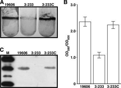
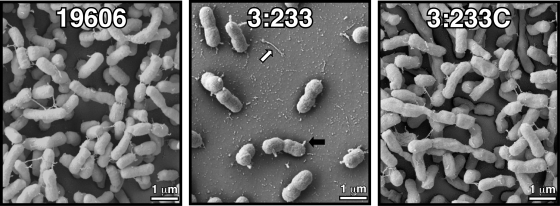
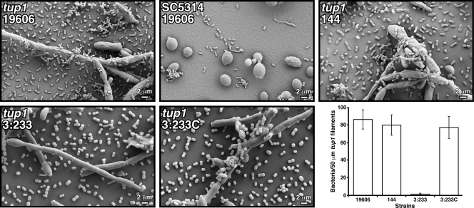
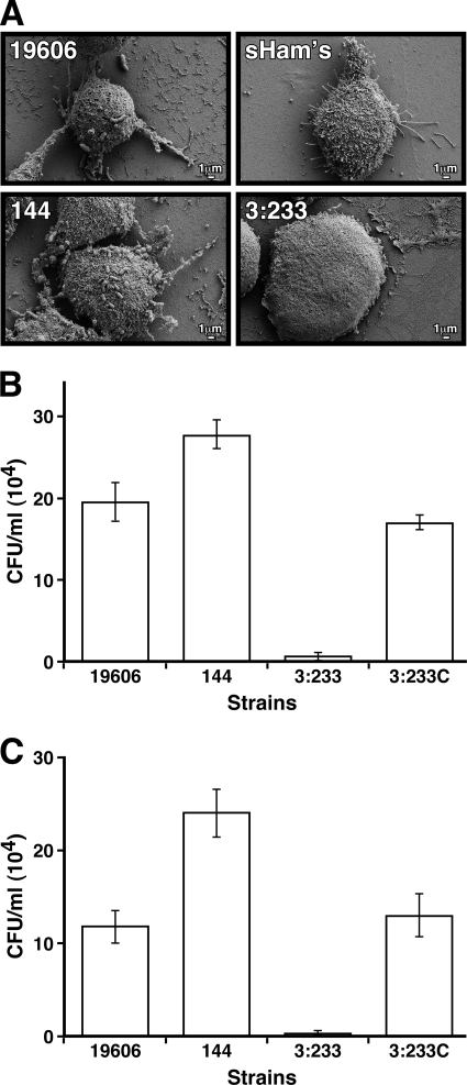
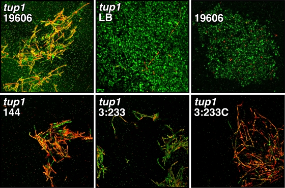
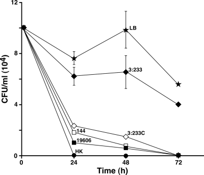
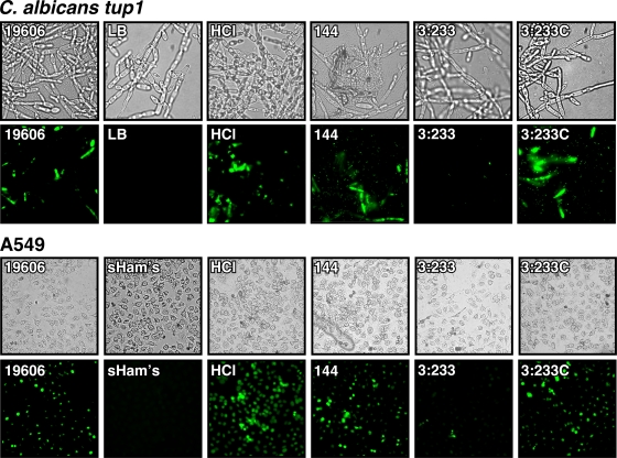
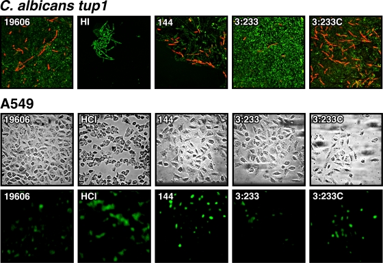
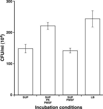
The ability of Acinetobacter baumannii to adhere to and persist on surfaces as biofilms could be central to its pathogenicity. The production of pili and a biofilm-associated protein and the expression of antibiotic resistance are needed for robust biofilm formation on abiotic and biotic surfaces. This multistep process also depends on the expression of transcriptional regulatory functions, some of which could sense nutrients available to cells. This report extends previous observations by showing that although outer membrane protein A (OmpA) of A. baumannii 19606 plays a partial role in the development of robust biofilms on plastic, it is essential for bacterial attachment to Candida albicans filaments and A549 human alveolar epithelial cells. In contrast to abiotic surfaces, the interaction with biotic surfaces is independent of the CsuA/BABCDE-mediated pili. The interaction of A. baumannii 19606 with fungal and epithelial cells also results in their apoptotic death, a response that depends on the direct contact of bacteria with these two types of eukaryotic cells. Furthermore, the bacterial adhesion phenotype correlates with the ability of bacteria to invade A549 epithelial cells. Interestingly, the killing activity of cell-free culture supernatants proved to be protease and temperature sensitive, suggesting that its cytotoxic activity is due to secreted proteins, some of which are different from OmpA.


Figures

FIG. 1.
FIG. 1.
Biofilm assays and detection of OmpA produced by A. baumannii 19606 derivatives. (A) Crystal violet staining of biofilms formed on 3-ml plastic tubes by parental strain 19606, the OmpA-deficient derivative 3:233, and ompA-complemented strain 3:233C. (B) Quantification of the biofilms shown in A. Error bars, 1 standard deviation (SD). OD580, optical density at 580 nm. (C) Western blotting of total membrane proteins (7 μg per lane) isolated from cells of the strains shown in A. Lane M, molecular mass markers. The top, middle, and bottom most-intense bands represent the mobilities of the 50-kDa, 37-kDa, and 25-kDa markers, respectively. After SDS-PAGE, proteins were blotted onto nitrocellulose and probed with anti-OmpA antiserum.
FIG. 2.
FIG. 2.
SEM analysis of bacterial biofilms formed on plastic surfaces at the liquid-air interface. The black and white arrows indicate cell protrusions and pili on the plastic surface, respectively.
FIG. 3.
FIG. 3.
SEM analysis of bacterial attachment to C. albicans cells. SC5314 yeast cells and tup1 strain filaments were coincubated with bacterial cells from parental strain 19606 or cells from the isogenic derivative 144, 3:233, or 3:233C. All micrographs were taken at a ×2,000 magnification. The bottom right represents the quantification of visible bacteria attached to 50 μm of tup1 strain filaments. Error bars, 1 SD.
FIG. 4.
FIG. 4.
Bacterial adhesion and invasion of A549 alveolar epithelial cells. (A) SEM analysis of A549 cells incubated in sterile supplemented Ham's tissue culture medium (sHam's) or in the presence of cells from parental strain 19606 or cells from the isogenic derivative 144, 3:233, or 3:233C. (B) Bacterial counts obtained after A549 cells were infected and lysed. (C) Bacterial counts obtained after A549 cells were infected, treated with gentamicin, and then lysed. Error bars, 1 SD.
FIG. 5.
FIG. 5.
LSCM of Live/Dead-stained tup1 strain filaments or SC5314 yeast cells incubated in sterile LB broth or in the presence of 19606 parental cells or cells from the isogenic derivative 144, 3:233, or 3:233C. Micrographs were taken at a ×400 magnification.
FIG. 6.
FIG. 6.
C. albicans viability assays. tup1 strain filaments were incubated in sterile LB broth or in the presence of bacterial cells from parental strain 19606 or the isogenic derivative 144, 3:233, or 3:233C. Heat-killed (HK) tup1 strain filaments were used as a negative control. The results represent three independent experiments performed under the same experimental conditions. Error bars, 1 SD.
FIG. 7.
FIG. 7.
TUNEL assays of C. albicans tup1 strain filaments and A549 monolayers cultured in sterile media (LB and supplemented Ham's medium [sHam's]) in the absence or in the presence of 2 M HCl (HCl), conditions which were used as negative and positive controls, respectively. The eukaryotic cells were also coincubated with bacterial cells from parental strain 19606 or with cells from the isogenic derivatives 144, 3:233, and 3:233C. Fungal filaments and A549 cells were imaged at ×600 and ×400 magnifications, respectively.
FIG. 8.
FIG. 8.
Killing of tup1 strain filaments and A549 cells by cell-free bacterial culture supernatants. (Top) LSCM of Live/Dead-stained tup1 strain filaments incubated with cell-free supernatants of cultures of strains 19606, 144, 3:233, and 3:233C grown overnight. HI, the 19606 culture supernatant was heat inactivated prior to addition to fungal filaments. Micrographs were taken at a ×400 magnification. (Bottom) TUNEL assays of A549 monolayers cultured in supplemented Ham's (sHam's) medium containing 2 M HCl (HCl) or cell-free supernatants of cultures of strains 19606, 144, 3:233, and 3:233C grown overnight. Fungal filaments and A549 cells were imaged at ×600 and ×400 magnifications, respectively.
FIG. 9.
FIG. 9.
Protease sensitivity of the cytotoxic factor(s) secreted by A. baumannii 19606. Fungal filaments were coincubated for 24 h with stationary-phase untreated 19606 cell-free supernatants (SUP) or cell-free supernatants that were treated with either proteinase K (PK) and PMSF or PMSF prior coincubation with tup1 strain filaments. Coincubation with sterile LB broth was used as a control. Error bars, 1 SD.

References

    1. Actis, L. A., S. A. Potter, and J. H. Crosa. 1985. Iron-regulated outer membrane protein OM2 of Vibrio anguillarum is encoded by virulence plasmid pJM1. J. Bacteriol. 161736-742. - PMC - PubMed
    1. Barcak, J. G., M. S. Chandler, R. J. Redfield, and J. F. Tomb. 1991. Genetic systems in Haemophilus influenzae. Methods Enzymol. 204321-432. - PubMed
    1. Barrios, A. F., R. Zuo, D. Ren, and T. K. Wood. 2006. Hha, YbaJ, and OmpA regulate Escherichia coli K12 biofilm formation and conjugation plasmids abolish motility. Biotechnol. Bioeng. 93188-200. - PubMed
    1. Bergogne-Berezin, E. 2001. The increasing role of Acinetobacter species as nosocomial pathogens. Curr. Infect. Dis. Rep. 3440-444. - PubMed
    1. Bouvet, P. J. M., and P. A. D. Grimont. 1986. Taxonomy of the genus Acinetobacter with the recognition of Acinetobacter baumannii sp. nov., Acinetobacter haemolyticus sp. nov., Acinetobacter johnsonii sp. nov., and Acinetobacter junii sp. nov. and emended descriptions of Acinetobacter calcoaceticus and Acinetobacter lwoffii. Int. J. Syst. Bacteriol. 36228-240.

Publication types

Substances