. 2009 Aug;29(15):4144-55.
doi: 10.1128/MCB.00380-09. Epub 2009 May 26.

Functional dissection of the human TNRC6 (GW182-related) family of proteins

Affiliations

Functional dissection of the human TNRC6 (GW182-related) family of proteins

David Baillat et al. Mol Cell Biol. 2009 Aug.

Abstract

Argonaute (Ago) proteins through their association with small RNAs perform a critical function in the effector step of RNA interference. The TNRC6 (trinucleotide repeat containing 6) family of proteins have been shown to stably associate with Agos in mammalian cells. Here, we describe the isolation and functional characterization of TNRC6B- and TNRC6C-containing complexes. We show that TNRC6B and TNRC6C proteins associate with all four human Agos which are already loaded with microRNAs. Detailed domain analysis of TNRC6B protein indicated that distinct domains of the protein are required for Ago binding and P-body localization. Functional analysis using reporter constructs responsive to TNRC6B tethered through an MS2-binding domain indicates that neither the Ago-binding nor the P-body localization domains are required for translational silencing. In contrast, the C-terminal domain containing the RNA recognition motif plays a critical role in the silencing mediated by the TNRC6B protein.


Figures

FIG. 1.
FIG. 1.
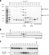
The TNRC6 family of proteins interacts with Argonaute proteins. (a) Purification of Ago1-associated protein complexes. (Left) Purification scheme. Flag-Ago1, Flag-tagged Ago1. (Right) Ago1-associated polypeptides. HEK293 cell lines stably expressing a Flag-tagged Ago1 protein were established. Polypeptides associated with Ago1 were immunopurified from S100 fractions from these cells using agarose beads conjugated with anti-Flag M2 monoclonal antibodies. After the polypeptides were washed and eluted, they were resolved by SDS-PAGE and silver stained. The parental HEK293 cell line was used as a control (mock) for the immunopurification. The positions of molecular mass markers are shown to the left of the gels. (b) Schematic alignment of TNRC6 family proteins and their D. melanogaster (dm) and C. elegans (ce) homologs. Gray blocks represent domains of higher amino acid sequence identity. The positions of domains I to IV, Q/P-rich domain (Q/P), RNA recognition motif (RRM), and ubiquitin-associated motif (UBA) are shown. The percent identity (percent similarity in parentheses) between the conserved domains of TNRC6C and Gawky is indicated. aa, amino acids. (c) Purification of TNRC6B- and TNRC6C-associated protein complexes. (Right) Purification of TNRC6B- and TNRC6C-associated polypeptides. H1299 cell lines stably expressing a Flag-tagged TNRC6C or TNRC6B protein were established. Polypeptides associated with TNRC6C or TNRC6B were immunopurified from S100 fractions from these cells using agarose beads conjugated to anti-Flag M2 monoclonal antibodies. After the polypeptides were washed and eluted, they were resolved by SDS-PAGE and silver stained. The positions of major degradation fragments of Flag-tagged TNRC6B (Flag-TNRC6B) and TNRC6C are indicated by asterisks besides the gels.
FIG. 2.
FIG. 2.
Analysis of the TNRC6B-associated complexes. (a) SDS-PAGE analysis of the TNRC6B-associated complex fractionated on a Superose 6 gel filtration column after silver staining. The Superose 6 fractions (fractions 14 to 38) are shown above the gel. The positions of molecular mass markers are indicated to the left of the gel. The positions of major contaminant polypeptides are indicated by asterisks. (b) The same fractions were analyzed by Western blotting with anti-Flag M2 and anti-Ago2 antibodies. (c) RISC activity after preincubation of the Superose 6 fractions with an siRNA trigger corresponding to the let-7 sequence.
FIG. 3.
FIG. 3.
Ago proteins interacting with TNRC6B and TNRC6C are already loaded with mature miRNAs and active. (a) Comparison of RISC activity between the TNRC6B- and Dicer-associated complexes with (+) or without (−) preincubation with a siRNA trigger (let-7). Flag-TNRC6B, Flag-tagged TNRC6B. (b) Northern blot analysis of TNRC6B- and TNRC6C-associated complexes. RNAs were extracted from the TNRC6B- and TNRC6C-associated complexes, precipitated, and resolved on a 15% acrylamide-7 M urea gel. After the RNAs were transferred from the gel to a membrane, the membranes were hybridized with 32P-labeled probes corresponding to the indicated miRNAs. The parental HEK293 cell line was used as a control (mock) for the immunopurification. Flag-IP, Flag immunoprecipitation. (c) Comparison of RISC activity corresponding to the let-7 miRNA between TNRC6B- and TNRC6C-associated complexes in the absence of any exogenous siRNA trigger. (d) Pre-miRNA processing assay. Dicer- and TNRC6B-associated complexes were immunopurified from stably expressing cells. Increasing amounts (in microliters) were tested for processing of pre-miR30a into mature miR30a. (e) Cells stably expressing TNRC6B were transfected with either 70-bp hairpins or siRNA corresponding to the luciferase sequence. After 24 h, the TNRC6B-associated complexes were purified by immunoaffinity, and their RISC activity was assayed against a luciferase probe (Luc probe) and a let7 probe. For controls, untransfected cells were used and the specificity was assessed by adding 2′-O-methyl (2′OMe) oligonucleotides complementary to the luciferase siRNA sequence. GL3 hp, luciferase hairpin. (f) Same as panel e except the luciferase hairpin was cotransfected with increasing amounts of a plasmid expressing the target sequence (pGL3-ctrl; Promega).
FIG. 4.
FIG. 4.
The glutamine/proline-rich domain localizes TNRC6B to P-bodies. (a) Schematic representation of the Flag-tagged TNRC6B deletion mutants used. The positions of domains I to IV, the Q/P-rich motif, and RRM are shown. aa, amino acids. (b) Cellular localization of TNRC6B. HeLa cells were transfected with the indicated constructs. After fixation and permeabilization, the cells were probed with anti-Flag M2 monoclonal antibodies (revealed with anti-mouse IgG antibodies coupled to Alexa Fluor 488) and with anti-DCP1a polyclonal antibodies (revealed with anti-rabbit IgG antibodies coupled to Alexa Fluor 568).
FIG. 5.
FIG. 5.
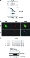
A single point mutation in domain I of TNRC6B abrogates Ago binding. (a) HEK293 cells were transfected with the different TNRC6B constructs described in the legend to Fig. 4a. Total cell extracts were immunoprecipitated with anti-Flag M2 antibodies. Immunoprecipitates were resolved by SDS-PAGE and analyzed by Western blotting with anti-Flag M2 antibodies and anti-Ago2 antibodies. Flag-IP, Flag immunoprecipitation. (b) HeLa cells were transfected with the indicated constructs. After fixation and permeabilization, the cells were probed with anti-Flag M2 monoclonal antibodies (revealed with anti-mouse IgG antibodies coupled to Alexa Fluor 488) and with anti-DCP1a polyclonal antibodies (revealed with anti-rabbit IgG antibodies coupled to Alexa Fluor 568). (c) (Top) Protein alignment for the highly conserved region in the Ago-binding domain of TNRC6B proteins from human (Homo sapiens [Hs]), zebra fish (Danio rerio [Dr]), and fruit fly (Drosophila melanogaster [Dm]) sources. (Bottom) Same as in panel b using the wild-type (WT) and W623A point mutant (mut) of full-length TNRC6B and D-I. ctrl, control.
FIG. 6.
FIG. 6.
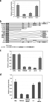
TNRC6B exerts a strong Ago-independent translational inhibition. (a) HEK293 cells were transfected with pGL3-MS2, pRL-CMV, and the various MS2 fusion constructs indicated. Twenty-four hours posttransfection, the cells were lysed, and firefly and Renilla luciferase activities were measured and plotted as the firefly luciferase/Renilla luciferase ratio. (b) Schematic representations of the TNRC6B-MS2 fusion constructs and the luciferase constructs used in this figure. The positions of MS2 domain, domains I to IV, Q/P-rich motif, and RRM are shown. aa, amino acids; SV40, simian virus 40; MBS x2, two copies of an MS2 binding site. (c and d) Same as in panel a. CTRL, control.
FIG. 7.
FIG. 7.
Translation inhibition exerted by the RRM-containing C-terminal domain of TNRC6B. (a) HEK293 cells were transfected with pGL3-MS2, pRL-CMV, and the various MS2 fusion constructs indicated. Twenty-four hours posttransfection, the cells were lysed, and firefly and Renilla luciferase activities were measured and plotted as the change in repression from the relative luciferase activity measured with the MS2 domain alone. (b) Cap-binding assay. HEK293 cells were transfected with Flag-tagged CBP20 or Flag-tagged ΔN1409. After 48 h, the cells were lysed, and Flag proteins were immunopurified. Eluted proteins were tested for cap binding using 7mGTP beads in the presence of specific (7mGpppG) or nonspecific (GpppG) competitor. Bound proteins were eluted with 7mGpppG and analyzed by Western blotting using anti-Flag antibodies. (c) (Top) Schematic representation of the EMCV internal ribosome entry site (IRES) luciferase construct. luc, luciferase; SV40, simian virus 40; MBS x2, two copies of an MS2 binding site. (Bottom) HEK293 cells were transfected with EMCV-luc-MS2 construct, pRL-CMV, and the MS2 fusion constructs indicated. Twenty-four hours posttransfection, the cells were lysed, and firefly and Renilla luciferase activities were measured and plotted as the firefly luciferase/Renilla luciferase ratio. (d) Same as in panel a but relative luciferase activity (firefly luciferase/Renilla luciferase) is plotted.

Similar articles

Cited by

References

    1. Bagga, S., J. Bracht, S. Hunter, K. Massirer, J. Holtz, R. Eachus, and A. E. Pasquinelli. 2005. Regulation by let-7 and lin-4 miRNAs results in target mRNA degradation. Cell 122553-563. - PubMed
    1. Bashkirov, V. I., H. Scherthan, J. A. Solinger, J. M. Buerstedde, and W. D. Heyer. 1997. A mouse cytoplasmic exoribonuclease (mXRN1p) with preference for G4 tetraplex substrates. J. Cell Biol. 136761-773. - PMC - PubMed
    1. Behlke, M. A., S. A. Dames, W. H. McDonald, K. L. Gould, E. J. Devor, and J. A. Walder. 2000. Use of high specific activity StarFire oligonucleotide probes to visualize low-abundance pre-mRNA splicing intermediates in S. pombe. BioTechniques 29892-897. - PubMed
    1. Behm-Ansmant, I., J. Rehwinkel, T. Doerks, A. Stark, P. Bork, and E. Izaurralde. 2006. mRNA degradation by miRNAs and GW182 requires both CCR4:NOT deadenylase and DCP1:DCP2 decapping complexes. Genes Dev. 201885-1898. - PMC - PubMed
    1. Carthew, R. W., and E. J. Sontheimer. 2009. Origins and mechanisms of miRNAs and siRNAs. Cell 136642-655. - PMC - PubMed

Publication types

MeSH terms