. 2009 Jun;2(6):572-80.
doi: 10.1158/1940-6207.CAPR-09-0001. Epub 2009 May 26.

A novel sulindac derivative that does not inhibit cyclooxygenases but potently inhibits colon tumor cell growth and induces apoptosis with antitumor activity

Affiliations

A novel sulindac derivative that does not inhibit cyclooxygenases but potently inhibits colon tumor cell growth and induces apoptosis with antitumor activity

Gary A Piazza et al. Cancer Prev Res (Phila). 2009 Jun.

Abstract

Nonsteroidal anti-inflammatory drugs such as sulindac have shown promising antineoplastic activity, although toxicity from cyclooxygenase (COX) inhibition and the suppression of prostaglandin synthesis limits their use for chemoprevention. Previous studies have concluded that the mechanism responsible for their antineoplastic activity may be COX independent. To selectively design out the COX inhibitory activity of sulindac sulfide (SS), in silico modeling studies were done that revealed the crucial role of the carboxylate moiety for COX-1 and COX-2 binding. These studies prompted the synthesis of a series of SS derivatives with carboxylate modifications that were screened for tumor cell growth and COX inhibitory activity. A SS amide (SSA) with a N,N-dimethylethyl amine substitution was found to lack COX-1 and COX-2 inhibitory activity, yet potently inhibit the growth of human colon tumor cell lines, HT-29, SW480, and HCT116 with IC(50) values of 2 to 5 micromol/L compared with 73 to 85 micromol/L for SS. The mechanism of growth inhibition involved the suppression of DNA synthesis and apoptosis induction. Oral administration of SSA was well-tolerated in mice and generated plasma levels that exceeded its in vitro IC(50) for tumor growth inhibition. In the human HT-29 colon tumor xenograft mouse model, SSA significantly inhibited tumor growth at a dosage of 250 mg/kg. Combined treatment of SSA with the chemotherapeutic drug, Camptosar, caused a more sustained suppression of tumor growth compared with Camptosar treatment alone. These results indicate that SSA has potential safety and efficacy advantages for colon cancer chemoprevention as well as utility for treating malignant disease if combined with chemotherapy.


Conflict of interest statement

Disclosure of Potential Conflicts of Interest

No potential conflicts of interest were disclosed.

Figures
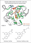
Fig. 1
Fig. 1
Molecular modeling of SS binding at the active site pocket of COX-1 or COX-2. Top, SS docked into the crystal structures of COX-1 and COX-2. Amino acids that participate in binding of SS to both COX-1 and COX-2 are illustrated through side chain heavy atoms and α carbons. Bottom, the two-dimensional chemical structure of SS and SSA.
Fig. 2
Fig. 2
Inhibition of colon tumor cell growth and DNA synthesis by SS and SSA. A and B, dose-dependent growth inhibitory activity of SS (A) and SSA (B) in human HT-29, HCT116, and SW480 colon tumor cells as measured by the CellTiterGlo assay described under “Materials and Methods.” C, the potency of SSA and SS, sulfone and sulfoxide to inhibit DNA synthesis in human HT-29 colon tumor cells. D, a time course experiment of SSA and SS to inhibit HT-29 tumor cell growth in which cell viability was measured using the CellTiterGlo assay. Columns, mean; bars, SE.
Fig. 3
Fig. 3
Apoptosis induction by SSA and SS in human HT-29 colon tumor cells. A and B, time- (A) and dose-dependent (B) induction of caspases 3 and 7 activities upon treatment with SSA measured by the Caspase-Glo 3/7 assay described under “Materials and Methods.” For time course studies, SSA was tested at a concentration of 25 µmol/L, whereas dose-response studies involved treatment for 5 h. C, a dose-dependent increase in DNA strand breaks by SSA and SS as measured by the TUNEL assay as described under “Materials and Methods.” Representative histograms (C) depict a dose-dependent increase in green fluorescence in each treatment group (gray) when compared with the vehicle control (black outline). Treatment values are significantly different than those for vehicle control with P value of ≤0.001 (†) or P value of ≤0.005 (*). Data represent mean ± SE.
Fig. 4
Fig. 4
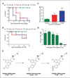
Tolerance and pharmacokinetics of SSA and sulindac in mice. A and B, the survival of mice treated with sulindac or SSA by gastric gavage, respectively. Mice were treated once daily with sulindac for 20 d and once daily with SSA for 17 d. C, plasma levels of SS, sulfone, and sulfoxide from mice 6 h after gastric gavage of sulindac at a dosage of 50 mg/kg. D, plasma levels of SSA from mice after various times after gastric gavage of SSA at a dosage of 200 mg/kg. Plasma levels of sulindac metabolites and SSA were measured as described under “Materials and Methods.” Columns, mean; bars, SE. The proposed metabolic pathway of SSA is shown in E.
Fig. 5
Fig. 5
In vivo antitumor efficacy of SSA and Camptosar alone and in combination as determined using the human HT-29 colon tumor xenograft mouse model. A, body weights of mice treated with vehicle or SSA administered twice daily by gastric gavage (day 1–55) at a dosage of 250 mg/kg. B, tumor growth of mice treated with vehicle or SSA at a dosage and schedule as described in A. C, tumor growth of mice treated with vehicle, SSA twice daily by gastric gavage (day 14–70) at a dosage of 250 mg/kg, Camptosar (day 14, 18, and 22) at a dosage of 40 mg/kg, i.v., or SSA and Camptosar (same dosages as single agent treatments). Treatment was initiated on the day of tumor implantation (day 0) for the study shown in A and B. For the study shown in C, treatment was initiated once the tumors reached a median size of 200 mg (day 14). Treatment values are significantly different than those for vehicle control with P value of ≤0.001 (**) or P ≤ 0.05 (*). Points, mean (A and B), or median (C) values; bars, SE.

Similar articles

Cited by

References

    1. Thun MJ, Henley SJ, Patrono C. Nonsteroidal anti-inflammatory drugs as anticancer agents: mechanistic, pharmacologic, and clinical issues. J Natl Cancer Inst. 2002;94:252–256. - PubMed
    1. Chan TA. Nonsteroidal anti-inflammatory drugs, apoptosis, and colon-cancer chemoprevention. Lancet Oncol. 2002;3:166–174. - PubMed
    1. Giardiello FM, Hamilton SR, Krush AJ. Treatment of colonic and rectal adenomas with sulindac in familial adenomatous polyposis. N Engl J Med. 1993;328:1313–1316. - PubMed
    1. Nugent KP, Farmer KC, Spigelman AD, Williams CB, Phillips RK. Randomized controlled trial of the effect of sulindac on duodenal and rectal polyposis and cell proliferation in patients with familial adenomatous polyposis. Br J Surg. 1993;80:1618–1619. - PubMed
    1. Matsuhashi N, Nakajima A, Fukushima Y, Yazaki Y, Oka T. Effects of sulindac on sporadic colorectal adenomatous polyps. Gut. 1997;40:344–349. - PMC - PubMed

Publication types

MeSH terms