. 2009 Sep;57(9):883-8.
doi: 10.1369/jhc.2009.953851. Epub 2009 May 26.

Immunoreactivity of glutamate in mouse retina inner segment of photoreceptors with in vivo cryotechnique

Affiliations

Immunoreactivity of glutamate in mouse retina inner segment of photoreceptors with in vivo cryotechnique

Nobuo Terada et al. J Histochem Cytochem. 2009 Sep.

Abstract

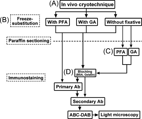
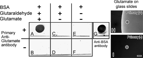
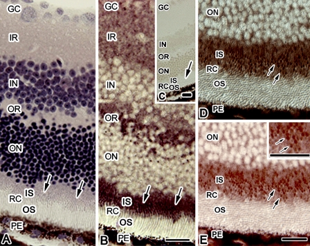
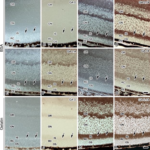
The purpose of this study was to clarify a previously controversial issue concerning glutamate (Glu) immunoreactivity (IR) in the inner segment (IS) of photoreceptors by using in vivo cryotechnique (IVCT) followed by freeze substitution (FS), which enabled us to analyze the cells and tissues reflecting living states. Eyeballs from anesthetized mice were directly frozen using IVCT. The frozen tissues were processed for FS fixation in acetone containing chemical fixatives, and embedded in paraffin. Deparaffinized sections were immunostained with an anti-Glu antibody. The strongest Glu-IR was obtained in the specimens prepared by FS with paraformaldehyde or a low concentration of glutaraldehyde, whereas no Glu-IR was obtained without the chemical fixatives. The Glu was immunolocalized in the IS, outer and inner plexiform and ganglion cell layers. Thus, the immunolocalization of Glu in the IS was clearly demonstrated using IVCT.


Figures

Figure 1
Figure 1
A flow diagram of the preparation steps for the mouse eyeball tissues, as prepared by the in vivo cryotechnique (IVCT) (A) and freeze-substitution (FS) fixation for the glutamate (Glu) immunostaining. During the FS, paraformaldehyde, glutaraldehyde, or no fixative was added to acetone (B). Some thin sections of eyeball tissues without the chemical fixative during FS were treated with paraformaldehyde or glutaraldehyde (C). Before immunoreaction of the primary antibody, a common blocking treatment with bovine serum albumin (BSA) or fish gelatin was performed on the sections (D). PFA, paraformaldehyde; GA, glutaraldehyde; Ab, antibody; ABC-DAB, horseradish-avidin-biotin complex and diaminobenzidine reactions.
Figure 2
Figure 2
Dot-blot analysis against variously prepared BSA with an anti-Glu antibody on nitrocellulose membranes (A–G). BSA-dotted membranes were treated with glutaraldehyde/Glu (A,B) or glutaraldehyde (C,D), or without treatment. BSA was assumed to be a binding dot pattern at the upper area of the line in each square. It was blocked with gelatin and incubated with (A,C,E) or without (B,D,F) the anti-Glu antibody. (G) Immunopositive control to show the binding of BSA as a dot pattern on the membrane with the anti-BSA antibody. Only after treatment with both glutaraldehyde and Glu (A), the immunoreactivity (IR) is obtained. (H,I) Glu-IR by attaching Glu to glass slides and immunostaining with (H) or without (I) the primary anti-Glu antibody (Primary) at higher magnification under a fluorescence microscope. Asterisks in (H) and (I) indicate the spot areas where the Glu was attached to glass slides. Bar = 50 μm.
Figure 3
Figure 3
Hematoxylin-eosin staining (A) and Glu immunostaining (B) images in adjacent sections of eyeball tissue obtained by IVCT followed by FS with paraformaldehyde. (C) Immunocontrol without the primary anti-Glu antibody. Arrows indicate the inner segment (IS) of photoreceptors. (D,E) Highly magnified view of diffraction interference contrast (DIC) (D) and Glu-immunostaining (E) images of a retina, in which photoreceptor cells in the outer segment (OS) and IS are obliquely sectioned. The Glu immunostaining is observed as tiny dot patterns (arrows in E) in the IS. Inset in E shows a higher magnified view of an area of the IS. GC, ganglion cell layer; IR and OR, inner and outer plexiform layers; IN and ON, inner and outer nuclear layers; RC, photoreceptor cell layer; PE, pigment epithelium. Bar = 20 μm.
Figure 4
Figure 4
DIC (A,C,E,G,I,K) and Glu immunostaining (B,D,F,H,J,L) images of the eyeball tissue sections treated with BSA (A–H) or fish gelatin (I–H) in samples with IVCT followed by FS with various concentrations of glutaraldehyde [0% (GA 0; A,B,I,J), 0.14% (GA 0.14; C,D,K,L), 1.4% (GA 1.4; E,F), and 14% (GA 14; G,H)]. Arrows indicate the IS of photoreceptors. Note that strong IR is detected with IVCT in eyeball tissues that were freeze-substituted with 0.14% glutaraldehyde, sectioned, and treated with BSA (D) or fish gelatin (L). IR and OR, inner and outer plexiform layers; IN and ON, inner and outer nuclear layers; RC, photoreceptor cell layer; IS and OS, inner and outer segments of RC; PE, pigment epithelium. Bar = 20 μm.

References

    1. Endo S, Ishiguro S, Tamai M (1999) Possible mechanism for the decrease of mitochondrial aspartate aminotransferase activity in ischemic and hypoxic rat retinas. Biochim Biophys Acta 1450:385–396 - PubMed
    1. Fletcher EL, Kalloniatis K (1996) Neurochemical architecture of the normal and degenerating rat retina. J Comp Neurol 376:343–360 - PubMed
    1. Kalloniatis M, Fletcher EL (1993) Immunocytochemical localization of the amino acid neurotransmitters in the chicken retina. J Comp Neurol 336:174–193 - PubMed
    1. Kalloniatis M, Marc RE, Murry RF (1996) Amino acid signatures in the primate retina. J Neurosci 16:6807–6829 - PMC - PubMed
    1. Marc RE, Liu WLS, Kalloniatis M, Raiguel SF, Van Haesendonck E (1990) Patterns of glutamate immunoreactivity in the goldfish retina. J Neurosci 10:4006–4034 - PMC - PubMed