Review
. 2009 Jul 1;103(4):655-63.
doi: 10.1002/bit.22361.

Hydrogels as extracellular matrix mimics for 3D cell culture

Affiliations
Review

Hydrogels as extracellular matrix mimics for 3D cell culture

Mark W Tibbitt et al. Biotechnol Bioeng. .

Abstract

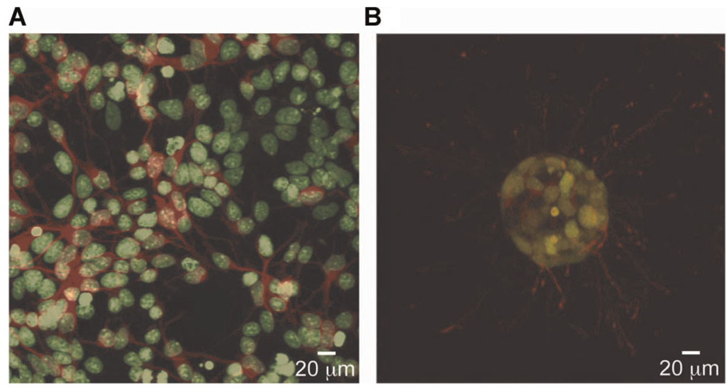
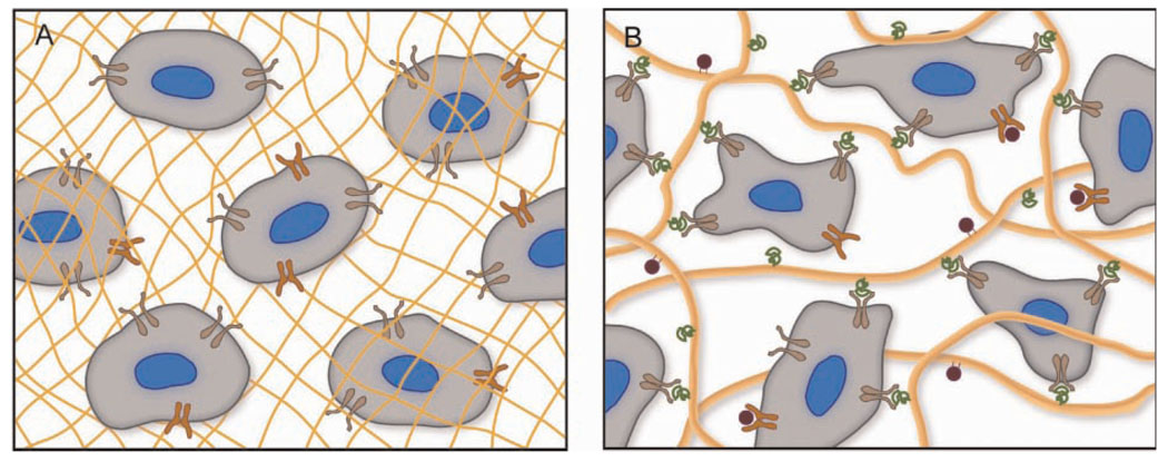
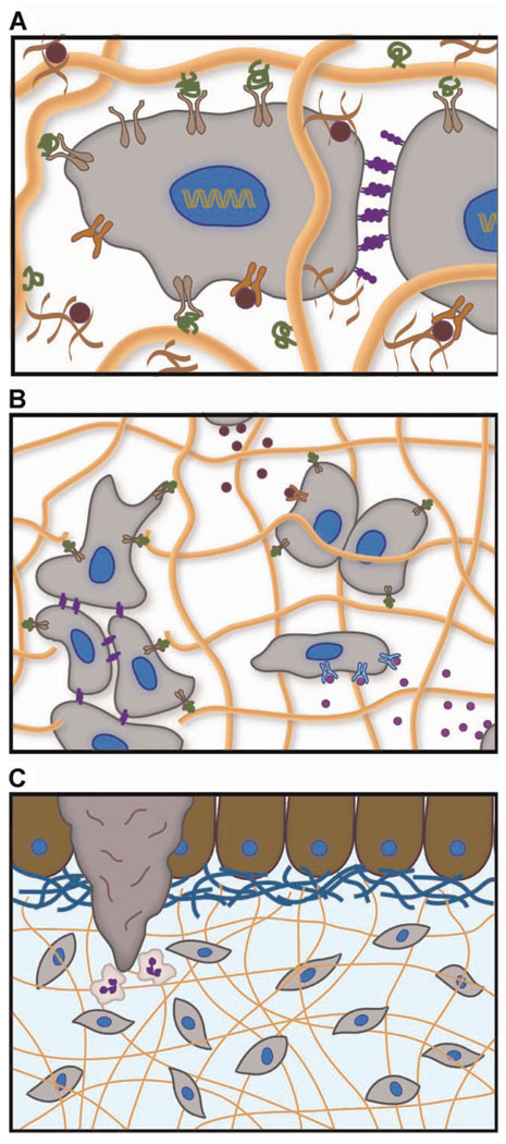
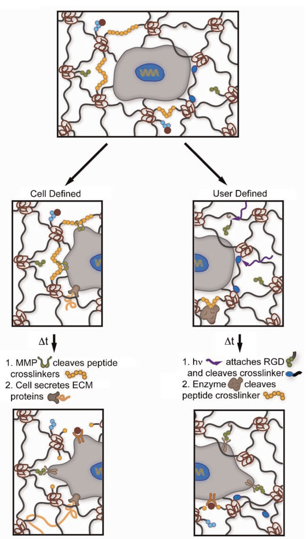
Methods for culturing mammalian cells ex vivo are increasingly needed to study cell and tissue physiology and to grow replacement tissue for regenerative medicine. Two-dimensional culture has been the paradigm for typical in vitro cell culture; however, it has been demonstrated that cells behave more natively when cultured in three-dimensional environments. Permissive, synthetic hydrogels and promoting, natural hydrogels have become popular as three-dimensional cell culture platforms; yet, both of these systems possess limitations. In this perspective, we discuss the use of both synthetic and natural hydrogels as scaffolds for three-dimensional cell culture as well as synthetic hydrogels that incorporate sophisticated biochemical and mechanical cues as mimics of the native extracellular matrix. Ultimately, advances in synthetic-biologic hydrogel hybrids are needed to provide robust platforms for investigating cell physiology and fabricating tissue outside of the organism.


Figures

Figure 1
Figure 1
Cells experience a drastically different environment between 2D and 3D culture. For instance, neural cells cultured in monolayer (A) are constrained to extend processes in the plane. Cell bodies are stained green and β-tubulin in axonal extensions is stained red. When cultured within hydrolytically degradable poly(ethylene glycol) based hydrogels (B) the same cells form neurospheres and extend processes isotropically in three dimensions. Images taken by M.J. Mahoney.
Figure 2
Figure 2
Permissive hydrogels (A) composed of synthetic polymers (yellow mesh) provide a 3D environment for culturing cells; however, they fail to activate integrins (brown) and other surface receptors (orange). The synthetic environment simply permits viability as cells remodel their surrounding microenvironment. On the other hand, promoting hydrogels (B) formed from naturally derived polymers present a myriad of integrin-binding sites (green) and growth factors (red) coordinated to the ECM (yellow fibers), which direct cell behavior through signaling cascades that are initiated by binding events with cell surface receptors.
Figure 3
Figure 3
The native ECM is the prototypical hydrogel that regulates cell function on many length scales. A: Integrin-binding with ECM proteins (green ligands and tan receptors), growth factor sequestration within proteoglycans (red), and cell–cell contact via cadherins (purple) occur on the scale of tens of nanometers to microns. B: Migration, which is critical in tissue regeneration, cancer metastasis, and wound healing, initiates on the scale of tens to hundreds of microns. Paracrine signaling that directs differentiation (pink growth factors) and proliferation (red growth factors) is also mediated on this length scale. C: Tissue homeostasis, development, and wound healing are regulated over hundreds of microns to centimeters. Here, we illustrate neutrophils being recruited to the site of a wound in the epithelium.
Figure 4
Figure 4
Synthetic–biologic hydrogels that incorporate several well-defined and orthogonal chemistries serve as robust ECM mimics for 3D cell culture. Depending on the application, it may be advantageous to incorporate cell- or user-defined regulation of the material properties to emulate the native dynamic environment. However, in many cases, synthetic hydrogels that incorporate both cell- and user-defined chemistries will be necessary. Here, we illustrate a cell cleaving MMP degradable crosslinks (yellow circles) that allow it to access sequestered growth factors (red) and integrin-binding sites, such as RGD (green circles). Ultimately, this cleavage allows cell motility and the deposition of ECM proteins (orange fiber). User-defined chemistries, such as photodegradable crosslinks (blue ellipses) and post-gelation attachment of RGD to the network backbone, afford facile control of the dynamic biochemical and biophysical properties of the gel, thereby directing cell attachment and motility. Further, exogenous application of enzymes (brown) can allow user-defined release of sequestered growth factors.

References

    1. Ashe HL, Briscoe J. The interpretation of morphogen gradients. Development. 2006;133(3):385–394. - PubMed
    1. Azab AK, Orkin B, Doviner V, Nissan A, Klein M, Srebnik M, Rubinstein A. Crosslinked chitosan implants as potential degradable devices for brachytherapy: In vitro and in vivo analysis. J Control Release. 2006;111(3):281–289. - PubMed
    1. Barralet JE, Wang L, Lawson M, Triffitt JT, Cooper PR, Shelton RM. Comparison of bone marrow cell growth on 2D and 3D alginate hydrogels. J Mater Sci Mater Med. 2005;16:515–519. - PubMed
    1. Birgersdotter A, Sandberg R, Ernberg I. Gene expression perturbation in vitro—A growing case for three-dimensional (3D) culture systems. Semin Cancer Biol. 2005;15(5):405–412. - PubMed
    1. Bryant SJ, Anseth KS. Hydrogel properties influence ECM production by chondrocytes photoencapsulated in poly(ethylene glycol) hydrogels. J Biomed Mater Res. 2002;59(1):63–72. - PubMed

Publication types

Substances

LinkOut - more resources