. 2009 Jul 15;115(14):3204-16.
doi: 10.1002/cncr.24397.

NV-128, a novel isoflavone derivative, induces caspase-independent cell death through the Akt/mammalian target of rapamycin pathway

Affiliations

NV-128, a novel isoflavone derivative, induces caspase-independent cell death through the Akt/mammalian target of rapamycin pathway

Ayesha B Alvero et al. Cancer. .

Abstract

Background: Resistance to apoptosis is 1 of the key events that confer chemoresistance and is mediated by the overexpression of antiapoptotic proteins, which inhibit caspase activation. The objective of this study was to evaluate whether the activation of an alternative, caspase-independent cell death pathway could promote death in chemoresistant ovarian cancer cells. The authors report the characterization of NV-128 as an inducer of cell death through a caspase-independent pathway.

Methods: Primary cultures of epithelial ovarian cancer (EOC) cells were treated with increasing concentration of NV-128, and the concentration that caused 50% growth inhibition (GI(50)) was determined using a proprietary assay. Apoptotic proteins were characterized by Western blot analyses, assays that measured caspase activity, immunohistochemistry, and flow cytometry. Protein-protein interactions were determined using immunoprecipitation. In vivo activity was measured in a xenograft mice model.

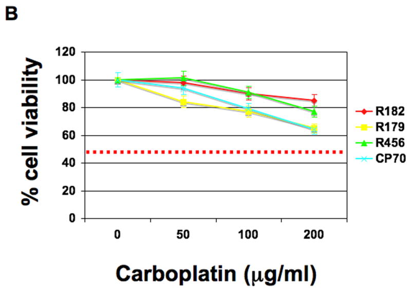
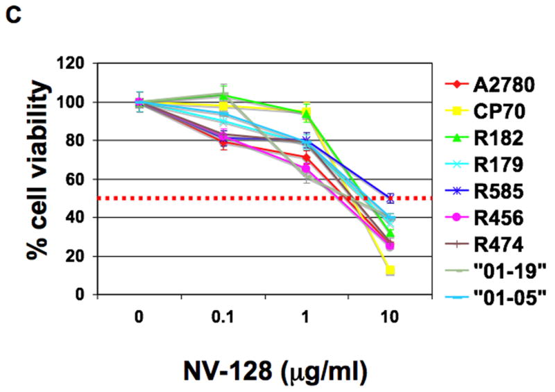
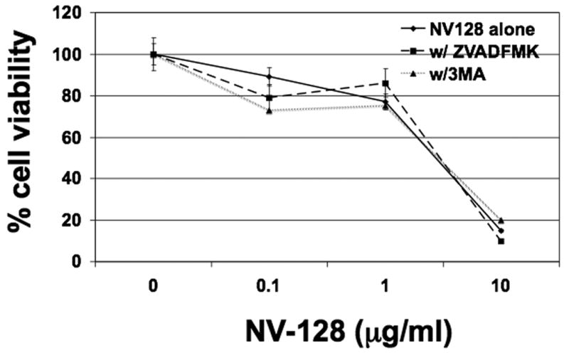
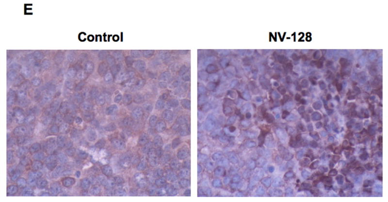
Results: NV-128 was able to induce significant cell death in both paclitaxel-resistant and carboplatin-resistant EOC cells with a GI(50) between 1 microg/mL and 5 microg/mL. Cell death was characterized by chromatin condensation but was caspase-independent. The activated pathway involved the down-regulation of phosphorylated AKT, phosphorylated mammalian target of rapamycin (mTOR), and phosphorylated ribosomal p70 S6 kinase, and the mitochondrial translocation of beclin-1 followed by nuclear translocation of endonuclease G.

Conclusions: The authors characterized a novel compound, NV-128, which inhibits mTOR and promotes caspase-independent cell death. The current results indicated that inhibition of mTOR may represent a relevant pathway for the induction of cell death in cells resistant to the classic caspase-dependent apoptosis. These findings demonstrate the possibility of using therapeutic drugs, such as NV-128, which may have beneficial effects in patients with chemoresistant ovarian cancer.


Figures

Figure 1
Figure 1. NV-128 decreases viability of Paclitaxel- and Carboplatin-resistant EOC cells
EOC cells were treated with increasing concentrations of (A) Paclitaxel, (B) Carboplatin, and (C) NV-128 for 24h and cell viability determined as described in the Materials and Methods section. Results shown are representative of three independent experiments. Note that GI50 was not reached for Paclitaxel and Carboplatin in lines that were considered resistant (dashed red line).
Figure 2
Figure 2. NV-128 does not induce caspase activation
(A) Caspase activity was measured in cell lysates obtained from EOC cells treated for 24h with increasing concentrations of NV-128 or 2μM Paclitaxel. Results shown are for R179. Similar results were observed in other lines tested. (B,C) EOC cells were treated with 10 μg/ml NV-128 for the indicated time and whole cells lysates were analyzed by western blot for XIAP and phospho-Akt (p-Akt). β-actin and total Akt (t-Akt) blots demonstrate even loading. (D) No-treatment control cells, and cells treated with NV-128 (10 μg/ml) for 24h, were stained with Hoechst and PI and analyzed by flow cytometry. Results shown are for R182. Similar results were observed in other lines tested.
Figure 3
Figure 3. NV-128 induces caspase- and autophagy-independent cell death
Cells were treated with increasing concentrations of NV-128 for 24h in the presence or absence of Z-VAD-FMK (20 μM) or 3-MA (10μM) and cell viability determined as described in the Materials and Methods section. Data shown are for R182, similar results were obtained with other lines analyzed.
Figure 4
Figure 4. NV-128 specifically down-regulates the mTOR pathway
(A) Levels of 8 phospho-proteins were measured in the lysates as described in the Materials and Methods section. Data shown are for R182, similar results were obtained with other lines analyzed. (B,C) EOC cells were treated with 10 μg/ml NV-128 for the indicated time and lysates were analyzed by western blot for phospho-mTOR (p-mTOR), phospho- p70S6 kinase (p-S6k) and LC3-II.
Figure 5
Figure 5. NV-128 induces intracellular vacuole formation and mitochondrial depolarization
Confocal microscope images of CP70 cells either unstimulated (A) or treated with 10 μg/ml NV-128 for 2h (B). Note the presence of intracellular vacuoles in B but not in A (red arrows). Fluorescent microscope images of cells stained with JC-1: unstimulated (C) or treated with 10 μg/ml NV-128 for 2h (D). Rounded arrow points to punctuate red staining in unstimulated cells; diamond-ending arrow points to a cell with some mitochondrial depolarization; and arrowhead points to a cell with bright green fluorescence suggesting most mitochondria have depolarized.
Figure 6
Figure 6. Quantitative analysis of mitochondrial depolarization using flow cytometry
EOC cells were treated with NV-128, stained with JC-1 dye, and analyzed by flow cytometry to quantify mitochondrial depolarization.
Figure 7
Figure 7. NV-128 induces Beclin-1 mitochondrial- and EndoG nuclear-translocation
(A) Western blot analysis of cell lysates and mitochondrial fractions prepared from EOC cells treated with 10 μg/ml NV-128. Total cell lysates were analyzed for full-length Bid and mitochondrial fractions were analyzed for Beclin-1 and Bax. β-actin and VDAC are shown as loading controls. (B) Western blot analysis of anti-Beclin immunoprecipitates, derived from mitochondrial fractions of NV-128 (10 μg/ml, 1h) treated EOC cells, probed with anti-Bcl-2 and anti-Bak. (C) Western blot analysis of nuclear fractions for AIF and EndoG. Topoisomerase I (Topo-I) is shown as loading control.
Figure 8
Figure 8. In vivo activity of NV-128
EOC tumors were established in s.c. in NCR nude mice and treatments were given as described in the Materials and Methods section. Tumor size was determined by caliper measurements. (A) EOC tumor proliferation kinetics; (B) excised tumors from representative mice dosed with vehicle control or NV-128; (C) representative mice from each treatment group – note that carbolatin-treated mice had significant weight loss; (D) Tumors from representative mice were lysed and analyzed by western blot analysis for p-S6K and t-S6k. (E) Paraffin-embedded sections of representative mouse tumors were analyzed for the localization of Endo by IHC.
Figure 9
Figure 9. In vivo toxicology studies
(A) Comparison of white blood cells (WBC WL×109/L), red blood cells (RBC × 1012/L), hematocrit (Hct), and hemoglobin (Hb g/L) levels in control and NV-128 treated animals. (B) Comparison of alkaline phosphatase (ALP U/L), alanine aminotransferase (ALT U/L), urea (mmol/L), and creatinine (Cre umol/L) in control and NV-128 treated animals.
Figure 10
Figure 10. Proposed mechanism of NV-128-induced cell death
(A) In unstimulated/healthy cells, the process of autophagy is inhibited by mTOR and anti-apoptotic proteins such as XIAP inhibit apoptosis. (B) NV-128 treatment induces down-regulation of p-mTOR and Beclin-1 mitochondrial translocation. Mitochondrial Beclin-1 inhibits Bcl2 and the resulting mitochondrial depolarization leads to EndoG nuclear translocation and chromatin condensation. Connection between p-mTOR and Beclin-1 mitochondrial translocation still remains to be determined.

References

    1. Schwartz PE. Current diagnosis and treatment modalities for ovarian cancer. Cancer Treat Res. 2002;107:99–118. - PubMed
    1. Igney FH, Krammer PH. Death and anti-death: tumour resistance to apoptosis. Nat Rev Cancer. 2002;2:277–288. - PubMed
    1. Edinger AL, Thompson CB. Death by design: apoptosis, necrosis and autophagy. Curr Opin Cell Biol. 2004;16:663–669. - PubMed
    1. Ashkenazi A, Dixit VM. Death receptors: signaling and modulation. Science. 1998;281:1305–1308. - PubMed
    1. Cory S, Adams JM. The Bcl2 family: regulators of the cellular life-or-death switch. Nat Rev Cancer. 2002;2:647–656. - PubMed

Publication types

MeSH terms