

A gene-alteration profile of human lung cancer cell lines
- PMID: 19472407
- PMCID: PMC2900846
- DOI: 10.1002/humu.21028
A gene-alteration profile of human lung cancer cell lines
Abstract
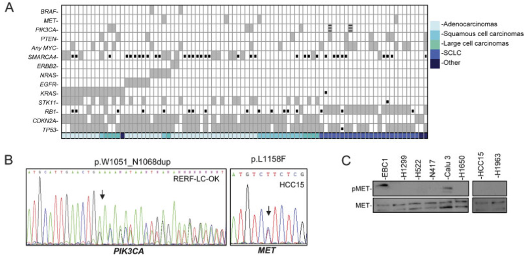
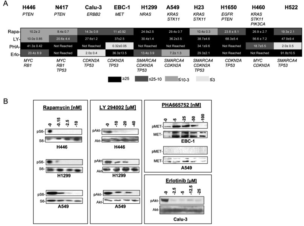
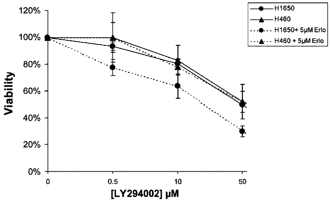
Aberrant proteins encoded from genes altered in tumors drive cancer development and may also be therapeutic targets. Here we derived a comprehensive gene-alteration profile of lung cancer cell lines. We tested 17 genes in a panel of 88 lung cancer cell lines and found the rates of alteration to be higher than previously thought. Nearly all cells feature inactivation at TP53 and CDKN2A or RB1, whereas BRAF, MET, ERBB2, and NRAS alterations were infrequent. A preferential accumulation of alterations among histopathological types and a mutually exclusive occurrence of alterations of CDKN2A and RB1 as well as of KRAS, epidermal growth factor receptor (EGFR), NRAS, and ERBB2 were seen. Moreover, in non-small-cell lung cancer (NSCLC), concomitant activation of signal transduction pathways known to converge in mammalian target of rapamycin (mTOR) was common. Cells with single activation of ERBB2, PTEN, or MET signaling showed greater sensitivity to cell-growth inhibition induced by erlotinib, LY294002, and PHA665752, respectively, than did cells featuring simultaneous activation of these pathways, underlining the need for combined therapeutic strategies in targeted cancer treatments. In conclusion, our gene-alteration landscape of lung cancer cell lines provides insights into how gene alterations accumulate and biological pathways interact in cancer.
Figures
References
-
- Angulo B, Suarez-Gauthier A, Lopez-Rios F, Medina PP, Conde E, Tang M, Soler G, Lopez-Encuentra A, Cigudosa JC, Sanchez-Cespedes M. Expression signatures in lung cancer show a profile for EGFR-mutant tumors and identifies selective PIK3CA overexpression by gene amplification. J Pathol. 2008;214:347–356. - PubMed
-
- Carpten JD, Faber AL, Horn C, Donoho GP, Briggs SL, Robbins CM, Hostetter G, Boguslawski S, Moses TY, Savage S, Uhlik M, Lin A, Du J, Qian YW, Zeckner DJ, Tucker-Kellogg G, Touchman J, Patel K, Mousses S, Bittner M, Schevitz R, Lai MH, Blanchard KL, Thomas JE. A transforming mutation in the pleckstrin homology domain of AKT1 in cancer. Nature. 2007;448:439–444. - PubMed
-
- Carretero J, Medina PP, Pio R, Montuenga LM, Sanchez-Cespedes M. Novel and natural knockout lung cancer cell lines for the LKB1/STK11 tumor suppressor gene. Oncogene. 2004;23:4037–4040. - PubMed
-
- Corradetti MN, Guan KL. Upstream of the mammalian target of rapamycin: do all roads pass through mTOR? Oncogene. 2006;25:6347–6360. - PubMed
-
- Engelman JA, Zejnullahu K, Mitsudomi T, Song Y, Hyland C, Park JO, Lindeman N, Gale CM, Zhao X, Christensen J, Kosaka T, Holmes AJ, Rogers AM, Cappuzzo F, Mok T, Lee C, Johnson BE, Cantley LC, Jänne PA. MET amplification leads to gefitinib resistance in lung cancer by activating ERBB3 signaling. Science. 2007;316:1039–1043. - PubMed
Publication types
MeSH terms
Grants and funding
LinkOut - more resources
Full Text Sources
Other Literature Sources
Medical
Research Materials
Miscellaneous
